Silent hypoxia is characterized by a significantly reduced oxygen saturation level with no associated breathing difficulties, at least in its beginning stages [1]. Because patients do not present with trouble breathing until the condition has progressed significantly, it can be difficult to detect [1]. Studies have recently reported that 20 to 40% of COVID-19 patients may experience silent hypoxia [1]. By the time medical teams have detected silent hypoxia, the patient may already have progressed into moderate-to-severe levels of infection, worsening their long-term recovery rates [2]. Consequently, medical practitioners must familiarize themselves with the warning signs of silent hypoxia to detect and treat it early.
There are many possible reasons why COVID-19 patients may develop silent hypoxia and, subsequently, experience a deterioration of their ability to breathe. Researchers have posited various hypotheses, from the virus’s impact on blood vessels contributing to impaired hypoxic vasoconstriction to changes to one’s respiratory system, perhaps via the expression of angiotensin-converting enzyme-2 receptors in the carotid bodies and nasal mucosa [1][3]. Unfortunately, while there are many possible theories concerning the development of this condition, research has not yet isolated the principal answer, making treatment more difficult [1].
While the underlying mechanisms guiding the development of silent hypoxia may remain uncertain, researchers have made progress in identifying certain predictors for the condition. In a retrospective cohort study, Alhusain and colleagues analyzed data from all of the COVID-19 patients who visited a hospital in Riyadh, Saudi Arabia over ten months [4]. Among the 195 patients who experienced silent hypoxia, the researchers identified no correlations between dyspnea and gender, age group, comorbidity, or body mass index [4]. This finding was partially consistent with another study by Okhuama et al., who found that age and diabetes were non-predictors of silent hypoxia in COVID-19 patients and suggested that more complex mechanisms were at play [5]. However, Alhusain et al. did find that fever, cough, and chronic cardiac disease were predictors of dyspnea, providing medical teams with some guidance in anticipating the condition [4].
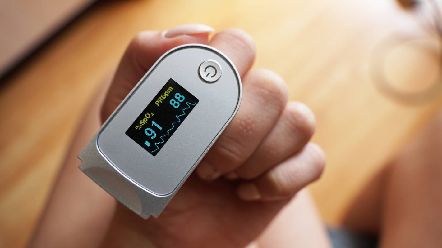
Medical teams can increase their chances of successfully identifying silent hypoxia by monitoring certain elements of the patient’s profile. Given that silent hypoxia patients may suffer from levels of pneumonia that are disproportionate to their COVID symptoms, practitioners should closely monitor patients’ pulse oximetry and arterial blood gas levels [5]. Bluish coloration can also indicate that a COVID-19 patient has silent hypoxia [6]. Before getting to the hospital setting, patients can also engage in detection practices themselves by regularly checking their blood saturation levels using a pulse oximeter or a smartphone [2]. A drop in oxygen saturation levels below 95% warrants contacting a health provider [2].
Ultimately, silent hypoxia is a pressing condition that can increase rates of intubation, mechanical ventilation, and even death among COVID-19 patients [2]. While medical teams still await more information about silent hypoxia, heeding the aforementioned indications can help treat pandemic patients and, moreover, suppress the adverse impacts of this ongoing pandemic.
References
[1] A. Rahman et al., “Silent hypoxia in COVID-19: pathomechanism and possible management strategy,” Molecular Biology Reports, vol. 48, no. 4, p. 3863-3869, April 2021. [Online]. Available: https://doi.org/10.1007/s11033-021-06358-1.
[2] J. Teo, “Early Detection of Silent Hypoxia in Covid-19 Pneumonia Using Smartphone Pulse Oximetry,” Journal of Medical Systems, vol. 44, no. 8, p. 1-2, June 2020. [Online]. Available: https://doi.org/10.1007/s10916-020-01587-6.
[3] T. Fuehner et al., “Silent Hypoxia in COVID-19: A Case Series,” Respiration, vol. 101, p. 376-380, November 2021. [Online]. Available: https://doi.org/10.1159/000520083.
[4] F. Alhusain et al., “Predictors and clinical outcomes of silent hypoxia in COVID-19 patients, a single-center retrospective cohort study,” Journal of Infection and Public Health, vol. 14, no. 11, p. 1595-1599, November 2021. [Online]. Available: https://doi.org/10.1016/j.jiph.2021.09.007.
[5] A. Okuhama et al., “Clinical and radiological findings of silent hypoxia among COVID-19 patients,” Journal of Infection and Chemotherapy, vol. 27, no. 10, p. 1536-1538, October 2021. [Online]. Available: https://doi.org/10.1016/j.jiac.2021.07.002.
[6] T. S. Simonson et al., “Silent hypoxaemia in COVID-19 patients,” The Journal of Physiology, vol. 599, no. 4, p. 1057-1065, December 2020. [Online]. Available: https://doi.org/10.1113/JP280769.