Categories
Uncategorized

Anesthesia Considerations for Thyroid Surgery 

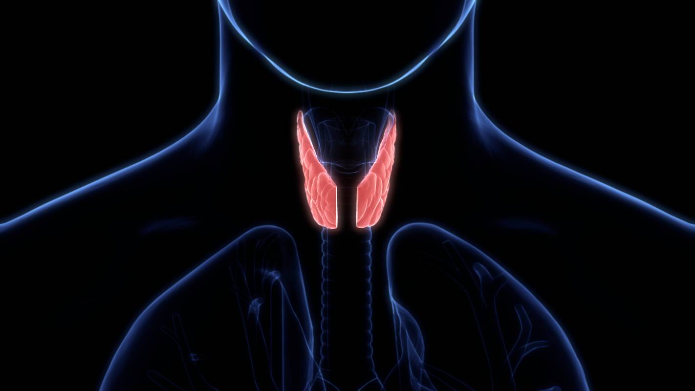
The thyroid is a gland located in the anterior region of the neck and is a critical part of the endocrine system.1 Specifically, the thyroid secretes the hormones T3 and T4, which increase the body’s basal metabolic rate, and calcitonin, which lowers blood calcium.2 Partial or complete removal of the thyroid in an operation known as a thyroidectomy may be recommended when cancer is detected or suspected to exist in the thyroid.3 Thyroid surgery may range from the simple removal of a small nodule from the thyroid to a more complicated operation to relieve the trachea from the pressure caused by a goiter (swelling of the neck resulting from enlargement of the thyroid), and the optimal anesthesia approach is affected by the type of surgery as well as patient factors.

Several different anesthesia options exist for thyroid surgery. Regional anesthesia has been shown to be effective and safe, though it is more often used for procedures on smaller scales.4 More common for a thyroidectomy, however, is general anesthesia. When tracheal compression is occurring, anesthesia can usually be administered without any impediments, however, a patient may be induced into the semi-supine or sitting position or, in more extreme cases, undergo fiberoptic intubation if needed.5 Pre-oxygenation with 100% oxygen should precede muscle relaxation for general anesthesia.

Total intravenous anesthesia, which is the use of intravenous agents only for the induction and maintenance of anesthesia, is commonly used during thyroid surgery.6 Propofol is among the most effective anesthetic agents, as it provides a rapid onset of anesthesia, does not have an extended recovery time, and has a low incidence of postoperative nausea.7 Opioids are often co-administered to potentiate their effect: the combination of propofol with the opioid remifentanil provides a short duration of action that can be easily targeted as well as adjusted for specific patient needs.8

There are several potential postoperative complications that anesthesiologists must be careful to detect and prevent from developing. One such complication is recurrent laryngeal nerve (RLN) palsy, a condition in which the recurrent laryngeal nerves that control the muscles of the larynx become injured, which can result in hoarseness, difficulty breathing, and physical exhaustion.9 While permanent nerve palsy occurs in less than 2% of patients undergoing thyroid surgery, temporary palsy has been reported in 3-10% of patients.10 The condition can be brought about by contusion to or clamping of the nerves during intubation and extubation, and, as such, anesthesiologists traditionally inspect the vocal cords immediately after extubation.5 Since this is often technically difficult, anesthesiologists can use a fiberscope or electrophysiological monitoring to prevent damage to the RLNs.11 An additional potential postoperative complication is postoperative hemorrhage, which can be avoided by maintaining hemostasis. Anesthesiologists typically maintain the patient’s intrathoracic pressure for 10-20 seconds to ensure that hemostasis has been achieved before the wound is closed.7

The parathyroid is a gland adjacent to the thyroid that maintains calcium homeostasis, and a parathyroidectomy, like a thyroidectomy, is a common surgical procedure for parathyroid swelling. In contrast to the thyroidectomy, however, local anesthesia is typically used for a parathyroidectomy, which may involve cervical nerve blocks.5 Nerve blocks may be deep or superficial, though no significant differences in postoperative pain and patient satisfaction have been found between the two types.

References 

1. Beynon, M. E. & Pinneri, K. An Overview of the Thyroid Gland and Thyroid-Related Deaths for the Forensic Pathologist. Acad. Forensic Pathol. 6, 217–236 (2016). 

2. How does the thyroid gland work? InformedHealth.org [Internet] (Institute for Quality and Efficiency in Health Care (IQWiG), 2018). 

3. Thyroid Surgery. American Thyroid Association https://www.thyroid.org/thyroid-surgery/

4. Hisham, A. N. & Aina, E. N. A reappraisal of thyroid surgery under local anaesthesia: back to the future? ANZ J. Surg. 72, 287–289 (2002). 

5. Malhotra, S. & Sodhi, V. Anaesthesia for thyroid and parathyroid surgery. Contin. Educ. Anaesth. Crit. Care Pain 7, 55–58 (2007). 

6. Total Intravenous Anaesthesia – an overview | ScienceDirect Topics. https://www.sciencedirect.com/topics/medicine-and-dentistry/total-intravenous-anaesthesia

7. Bacuzzi, A. et al. Anaesthesia for thyroid surgery: Perioperative management. Int. J. Surg. 6, S82–S85 (2008). 

8. Lentschener, C. et al. Remifentanil-propofol vs. sufentanil-propofol: optimal combinations in clinical anesthesia. Acta Anaesthesiol. Scand. 47, 84–89 (2003). 

9. Paquette, C. M., Manos, D. C. & Psooy, B. J. Unilateral Vocal Cord Paralysis: A Review of CT Findings, Mediastinal Causes, and the Course of the Recurrent Laryngeal Nerves. RadioGraphics 32, 721–740 (2012). 

10. Rafiq, M., Al-Zoraigi, U., Alzahrani, S. & Alabdulkarim, Y. A Case of Transient Local Anesthetic Induced Bilateral Vocal Cord Palsy. Case Rep. Surg. 2015, 379258 (2015). 

11. Tanigawa, K., Inoue, Y. & Iwata, S. Protection of recurrent laryngeal nerve during neck surgery: a new combination of neutracer, laryngeal mask airway, and fiberoptic bronchoscope. Anesthesiology 74, 966–967 (1991). 

Categories
Uncategorized

Antibodies After COVID-19 Infection

The COVID-19 pandemic, caused by the virus SARS-CoV-2, is still a global problem. Research on protection due to antibodies after COVID-19 infection (and vaccination) can contribute to clinical knowledge and more informed public health strategies.

Antibodies, also called immunoglobulins (Ig), are created by the plasma cells (mature B cells) of the immune system. The antibodies recognize and bind to a specific pattern or structure, called the antigen. Antibodies prevent harm by neutralizing a pathogen’s ability to enter cells or by marking the pathogen for attack by specialized immune cells (Durani, 2014). After a first infection, the body will keep some of the plasma cells as memory B cells, so that if the pathogen is encountered again, the antibody response will be quicker and more powerful (Cagiga et al, 2021).

The body makes five different types of antibodies: IgG, IgM, IgA, IgE and IgD. IgG is the most common antibody type and is a smaller protein that is found throughout the body. IgM is the first antibody type made upon infection and is found primarily in blood and lymph fluid. IgA is found mostly in mucous membranes, including in the respiratory tracts. IgE is mostly seen in allergic reactions, and IgD exists in only small amounts and its purpose is not understood (Durani, 2014). IgG, IgM and IgA are the types of antibodies that are relevant to SARS-CoV-2 infection.

For SARS-CoV-2, the antibody can attach to an antigen from either the virus’s spike glycoprotein (S antibodies) or nucleocapsid protein (N antibodies). The virus’s spike protein contains the receptor binding domain (RBD) that the virus uses to enter host cells; S antibodies against the RBD are more likely to be neutralizing (Cagiga et al, 2021). A neutralizing antibody prevents the virus from entering the host cell and reproducing. In comparison to S antibodies, N antibodies have not been found to provide protection against infection (Cagiga et al, 2021).

In response to COVID-19 infection, IgM antibodies are produced first throughout the body; it reacts strongly to antigens (high avidity) and ultimately represents 10% of the serum antibodies. IgG appears later and has high capacity for neutralization (Zhu et al, 2021). IgG and IgA are also produced locally from cells in the airway. IgA from the airway has been seen to peak at high levels early in infection and then begin to decline. Airway IgG and IgA levels waned significantly by 3 months post-infection in a study of 147 patients (Cagiga et al, 2021). Systemic IgM levels also declined significantly by the third month, and systemic IgG levels were observed to slightly decline (Zhu et al, 2021).

Despite declining levels, a high percentage of patients were still seropositive for IgG seven months after infection; in other words, they still had significant IgG reaction against a SARS-CoV-2 challenge. IgG antibodies after COVID-19 infection may stay in the body for up to two years (Zhu et al, 2021). It was also found that around 25% of patients still were seropositive for IgM after 6 to 9 months. However, all antibodies were found to decrease in neutralizing ability over time (Zhu et al, 2021).

In addition to antibody type, levels, and neutralizing ability, antibody avidity also plays a role in effectiveness. Avidity is how strongly the antibody binds to its antigen. With SARS-CoV-2, antibody avidity increased three months after infection when antibody levels started to decline, in a process called avidity maturation. High avidity antibodies may be associated with a lower risk of reinfection (Löfström et al, 2021).

Antibodies after recovery from COVID-19 were seen to vary based on disease severity. Patients recovering from a severe or critical disease were seropositive for IgG and IgM after approximately 7 months at a significantly higher rate than patients with asymptomatic infection (Zhu et al, 2021). Patients with low viral loads in the respiratory tract also may not develop antibodies at all (non-seroconversion) (Liu et al, 2021).

Age also impacted antibodies after recovery. Kids and adolescents were found to have the lowest rates of seropositivity for IgG and IgM, with adults under 60 having the highest rates and adults older than 60 falling in between. Luckily, children were more likely to have a less violent immune response and only mild symptoms, if any at all (Liu et al, 2021).

References 

Cagigi A, Yu M, Österberg B, et al. Airway antibodies emerge according to COVID-19 severity and wane rapidly but reappear after SARS-CoV-2 vaccination. JCI Insight. 2021;6(22):e151463. Published 2021 Nov 22. doi:10.1172/jci.insight.151463 

Löfström E, Eringfält A, Kötz A, et al. Dynamics of IgG-avidity and antibody levels after Covid-19. J Clin Virol. 2021;144:104986. doi:10.1016/j.jcv.2021.104986 

Liu W, Russell RM, Bibollet-Ruche F, et al. Predictors of Nonseroconversion after SARS-CoV-2 Infection. Emerg Infect Dis. 2021;27(9):2454-2458. doi:10.3201/eid2709.211042. 

Durani Y. Blood Test: Immunoglobulins (IgA, IgG, IgM). Rady Children’s Hospital San Diego- rchsd.org. 2014. https://www.rchsd.org/health-articles/blood-test-immunoglobulins-iga-igg-igm/

Zhu L, Xu X, Zhu B, et al. Kinetics of SARS-CoV-2 Specific and Neutralizing Antibodies over Seven Months after Symptom Onset in COVID-19 Patients. Microbiol Spectr. 2021;9(2):e0059021. doi:10.1128/Spectrum.00590-21

Categories
Uncategorized

Controversy Over “No Surprises Act” Implementation

Last December, Congress passed the “No Surprises Act” to shield patients from unexpected bills incurred when they receive medical treatment from providers outside their insurance networks. The bipartisan bill was applauded by groups like the American Hospitals Association,1 as well as consumers frustrated with the long history of “balance billing” in the United States — a process in which out-of-network providers bill individuals for the charges not covered by their insurance plans. The “No Surprises Act” attempts to remove patients from the center of these conflicts by establishing an independent dispute resolution process between insurers and providers.2 But as Congress finalizes the details of the implementation of the No Surprises Act, many providers are protesting new guidelines they argue will benefit insurers at the expense of patients and providers.3 

Patients often encounter surprise bills in emergency settings, when they go to (or are taken to) a facility without consideration of their insurance plan due to a focus on quickly receiving definite care. A 2017 study found that patients who received surprise bills for emergency care paid ten times as much as those who did not, and that roughly 18 percent of emergency visits resulted in at least one out-of-network bill.4 But surprise bills can also appear when patients go to an in-network facility but receive care from an out-of-network provider, such as an anesthesiologist, surgical assistant, or ambulatory service.2 Roughly one in five privately insured patients undergoing an elective surgery at in-network hospitals received such a bill, according to a 2020 study. This was often attributed to anesthesiology expenses, with an average out-of-network bill of $1,219 in the study.4  

According to a new rule of the No Surprises Act, a particular benchmark — the qualifying payment amount (QPA) — should serve as a “starting point” in making payment determinations, as this number is “generally the plan or issuer’s median contracted rate for the same or similar service in the specific geographic area,” according to the Centers for Medicare and Medicaid Services (CMS).2 Parties can submit additional information if they wish to make an offer that deviates from the QPA-based offer, but it “must clearly demonstrate that the value of the item or service is materially different from the QPA,” according to the CMS.2  

Now, members of Congress are arguing that prioritizing the QPA over other considerations (physician’s training, quality of outcomes, local market share of the parties involved, etc.), will give insurers in upper hand in the dispute resolution process. Relying on the QPA may incentivize insurers to set artificially low payment rates, putting pressure on small practices, like anesthesiology practices, and limiting patients’ access to care. Over 150 lawmakers — nearly half of them Democrats, and some of them doctors themselves — signed a letter citing their opposition to the new rule.3 

Also speaking out against the rule are leading physician societies, including the American Society of Anesthesiologists (ASA). These societies argue that the QPA is calculated by the insurance companies “without meaningful oversight or transparency,” and therefore can be manipulated in their favor without reflecting actual payment rates, thereby undermining the spirit of the legislation, which emphasizes information sharing and the equal consideration of multiple factors.For this reason, some health care experts have emphasized that “rule makers should prioritize, strengthen, and highlight full historical context for arbiters and emphasize this information-sharing provision in final rulemaking for the arbitration process.”6 

Already, the ASA is protesting actions believed to be driven by insurers’ desires to maintain the upper hand in forthcoming disputes. According to the ASA, Blue Cross Blue Shield of North Carolina threatened in a letter to anesthesiology and physician practices that their contract and in-network status would be terminated unless they immediately agreed to payment reductions ranging from 10% to over 30%, with the No Surprises Act cited as driving the reduction. To many, this shows how the new rule may allow insurance companies to leverage their market power to prioritize their finances, pushing providers out of insurance networks or forcing them to accept lower rates along the way.7 It may particularly harm networks in rural and underserved areas by incentivizing insurers to push down the rates they pay to in-network providers.3 

Still, in some areas, a united group of providers may be stronger than the insurers in the market, allowing them to take the upper hand. The Congressional Budget Office also reports that patients may enjoy lower premiums, reduced by an estimated 1%, as a result of the act.3 If the act is passed with these provisions, it ultimately remains to be seen what effect it will have. 

References 

  1. Detailed summary of No Surprises Act. (2021, January 14). American Hospitals Association. https://www.aha.org/advisory/2021-01-14-detailed-summary-no-surprises-act 
  1. Requirements Related to Surprise Billing; Part II interim final rule with comment period. (2021, September 30). Centers for Medicare and Medicaid Services. https://www.cms.gov/newsroom/fact-sheets/requirements-related-surprise-billing-part-ii-interim-final-rule-comment-period 
  1. McAuliff, M. (2021, November 17). Congressional doctors lead bipartisan revolt over policy on surprise medical bills. Kaiser Health News. https://khn.org/news/article/surprise-medical-bills-policy-congressional-doctors-backlash/ 
  1. Office of the Assistant Secretary for Planning and Evaluation. (2021, November 22). Evidence on Surprise Billing: Protecting Consumers with the No Surprises Act. U.S. Department of Health and Human Services. https://aspe.hhs.gov/sites/default/files/documents/acfa063998d25b3b4eb82ae159163575/no-surprises-act-brief.pdf 
  1. Nation’s frontline physicians denounce regulators’ implementation of key rule in No Surprises Act. (2021, October 1). American Society of Anesthesiologists. https://www.asahq.org/about-asa/newsroom/news-releases/2021/10/nations-frontline-physicians-denounce-regulators-implementation-of–key-rule-in-no-surprises-act 
  1. Koski-Vacirca, R., & Venkatesh, A. (2021, November 2). Rulemaking for health care affordability: Implementing the No Surprises Act. Health Affairs. https://www.healthaffairs.org/do/10.1377/hblog20211028.424036/full/  
  1. Lagasse, J. (2021, November 23). American Society of Anesthesiologists accuses BCBSNC of abusing No Surprises Act. Healthcare Finance News. https://www.healthcarefinancenews.com/news/american-society-anesthesiologists-accuses-bcbs-north-carolina-abusing-no-surprises-act 
Categories
Uncategorized

IV Ibuprofen for Analgesia

Ibuprofen is a non-steroidal anti-inflammatory drug (NSAID). Although most familiar in its oral form, ibuprofen can also be administered as an intravenous (IV) medication for more rapid effects. IV ibuprofen is approved in the US as an analgesic (pain reliever) and antipyretic (fever reducer) (Southworth and Sellers, 2020).

Ibuprofen’s main action within the body is the inhibition of prostaglandin synthesis. Prostaglandins are pro-inflammatory chemical signals that increase blood vessel permeability, recruit immune cells and promote edema (fluid accumulation). They also make pain receptors more sensitive to stimulation (Mazaleuskaya et al, 2015). Additionally, a prostaglandin synthesized in the hypothalamus (a region of the brain that works to maintain body homeostasis), PGE2, is a mediator of fever (Mazaleuskaya et al, 2015). Ibuprofen blocks prostaglandin synthesis by inhibiting the cyclooxygenas enzymes COX1 and COX2; it thus has anti-inflammatory, analgesic and antipyretic action (Mazaleuskaya et al, 2015). Additional pathways through which ibuprofen may act are still being explored. One interesting theory is that ibuprofen may be able to activate the brain’s cannabinoid receptors, which would decrease the perception of pain (Mazaleuskaya et al, 2015).

IV ibuprofen is often used with opioid medication as a multi-modal treatment plan for pain after surgery. Opioids are very strong pain relievers; however, they have a high risk of dependence and misuse, as well as other adverse effects such as medication interactions (Southworth and Sellers, 2020). They also do not address any underlying cause of pain, whereas NSAIDs are able to decrease inflammation (Southworth and Sellers, 2020). IV NSAIDs ketorolac and diclofenac are also approved in the US for pain relief but ibuprofen is the only IV NSAID currently approved for both pain relief and fever reduction (Southworth and Sellers, 2020).

IV ibuprofen has been shown to reduce the quantity of opioids required by a patient when compared to a placebo and other drugs like acetaminophen. Its use decreases patients’ subjective levels of pain as well as measurable levels of stress hormones in the postoperative period (Southworth and Sellers, 2020). Studies report that ibuprofen is well tolerated by patients and that there is no statistically significant increase in severe adverse events when compared with a placebo. It is also well-tolerated as a rapid infusion, given over 5-10 minutes (Southworth and Sellers, 2020).

Although generally considered safe, NSAIDs, including ibuprofen, have some side effects related to the inhibition of COX1 and COX2, which contribute to other processes throughout the body besides prostaglandin synthesis. Inhibition of COX1 is related to gastrointestinal (GI) symptoms and long-term inhibition of COX2 may have cardiovascular risks (Southworth et al, 2015). Ibuprofen is not selective for either COX1 or COX2, as compared with ketorolac which is more selective for COX1 and diclofenac which is more selective for COX2. Ibuprofen thus may have a more favorable risk profile in terms of GI and cardiovascular complications (Southworth et al, 2015).

IV ibuprofen may also have impacts on the effectiveness of other medications that patients take concurrently. Low dose aspirin, used for antiplatelet therapy to prevent stroke and heart attack, may be less effective since ibuprofen can block platelet COX1. This can also increase bleeding risk if patients are being given other anticoagulants or selective serotonin reuptake inhibitors (SSRI’s) (Mazaleuskaya et al, 2015). Ibuprofen may also decrease effectiveness of antihypertensive drugs, since the inhibition of prostaglandins in the kidney tubules causes the blood vessels to constrict, a mechanism of hypertension (Mazaleuskaya et al, 2015). Finally, patients taking lithium need to have their serum lithium levels monitored, since NSAIDS reduce lithium clearance from the body and can lead to toxic levels in the blood (Mazaleuskaya et al, 2015).

References 

Mazaleuskaya LL, Theken KN, Gong L, et al. PharmGKB summary: ibuprofen pathways. Pharmacogenet Genomics. 2015;25(2):96-106. doi:10.1097/FPC.0000000000000113 

Southworth SR, Sellers JA. Narrative Summary of Recently Published Literature on Intravenous Ibuprofen. Clin Ther. 2020;42(7):1210-1221. doi:10.1016/j.clinthera.2020.05.004 

Southworth SR, Woodward EJ, Peng A, Rock AD. An integrated safety analysis of intravenous ibuprofen (Caldolor(®)) in adults. J Pain Res. 2015;8:753-765. Published 2015 Oct 23. doi:10.2147/JPR.S93547 

Categories
Uncategorized

VR for Pediatric Medical Procedures

Originally developed for the entertainment industry as the next generation of video gaming systems, “virtual reality” (VR) presents exciting applications for the medical field. With a headset or “head-mounted display” (HMD), headphones equipped with noise reduction, a “rumble pad,” and joystick device, users are immersed into a virtual environment, complete with virtual objects, characters, and scenes that appear real. Using built-in head-tracking programs, HMD systems are able to follow the movements of the user and adjust the graphics accordingly, giving them the illusion of being completely surrounded by the virtual environment [1]. Unlike the two-dimensional experiences of playing a video game or watching a movie, VR systems like Oculus Rift and Samsung Gear employ multimodal stimuli to create immersive, distinct virtual environments that submerge the user into a different world. Despite its origin as the next form of entertainment, VR technology has exciting potential in the medical field, especially in the context of pediatric medical procedures.

In recent years, VR has been utilized in a variety of clinical settings, such as in cancer care, mental health treatment, and pain management [2, 3]. For example, using VR as a form of exposure therapy was shown to significantly decrease symptoms of post-traumatic stress disorder (PTSD) in veterans [4] and reduce severe phobias in patients with panic disorders [5]. Moreover, many physicians and researchers have begun to utilize VR in extremely painful or frightening situations, such as in burn care, and routine medical procedures, like IV placement, especially in pediatric patients, due to the theory that VR engages parts of the brain involved in pain perception and takes the user’s attention away from pain, leading to a perceived reduction in pain [6, 7]. One recent study measured changes in pain ratings in patients suffering from pain during hospitalization who either engaged in VR three times a day for 10 minutes per session or were instructed to watch guided meditation or yoga videos for the same intervals. The decrease in pain ratings for the VR group was significantly larger than for the control group [6]. VR combined with analgesia also significantly decreases pain and anxiety in patients undergoing burn debridement compared to analgesia alone [8].

Pediatric patients are particularly likely to experience anxiety, fear, and/or treatment non-adherence surrounding medical care due to the discomfort associated with routine procedures , and therefore may benefit from effective VR innovations [9]. In children, VR is particularly effective at reducing pain and anxiety — according to a large, randomized control trial, pediatric patients undergoing painful and distressing procedures such as intravenous catheter placement and blood draws who played a VR game during the procedure had significantly lower post-procedure pain and anxiety compared to patients who received only standard care and distraction techniques (i.e., coloring or listening to music) [9].

However, even though VR is one of the most effective methods of reducing pain and anxiety in pediatric patients, it is also one of the least commonly used [9]. Many experts recommend implementing VR in pediatric procedures in the future, especially as VR systems continue to become more cost-effective and accessible to healthcare providers [7].

References 

  1. Cipresso, P., Giglioli, I., Raya, M., and Riva, G. (2018). The past, present, and future of virtual and augmented reality research: a network and cluster analysis of the literature. Frontiers in Psychology, vol. 9. DOI: 10.3389/fpsyg.2018.02086. 
  1. Rothbaum, B., Hodges, L., and Kooper, R. (1997). Virtual reality exposure therapy. Journal of Psychotherapy Practice and Research, vol. 6. URL: https://www.ncbi.nlm.nih.gov/pmc/articles/PMC3330462/.  
  1. Morris, L., Louw, Q., and Grimmer-Somers, K. (2009). The effectiveness of virtual reality on reducing pain and anxiety in burn injury patients: a systematic review. The Clinical Journal of Pain, vol. 25. DOI: 10.1097/AJP.0b013e3181aaa909.  
  1. Rizzo, A., Difede, J., Rothbaum, B., Johnston, S., McLay, N., Reger, G., Gahm, G., Parsons, T., Graap, K., and Pair, J. (2009). VR PTSD exposure therapy results with active OIF/OEF combatants. Studies in Health Technology and Informatics, vol. 142. URL: https://pubmed.ncbi.nlm.nih.gov/19377167/.  
  1. Da Costa, R., de Carvalho, M., Ribero, P., and Nardi, A. (2018). Virtual reality exposure therapy for fear of driving: analysis of clinical characteristics, physiological response, and sense of presence. Brazilian Journal of Psychiatry, vol. 40. DOI: 10.1590/1516-4446-2017-2270.  
  1. Spiegel, B., Fuller, G., Lopez, M., Dupuy, T., Noah, B., Howard, A., Albert, M., Tashjian, V., Lam, R., Ahn, J., Dailey, F., Rosen, T., et al. (2019). Virtual reality for management of pain in hospitalized patients: a randomized comparative effectiveness trial. PLOS One, vol. 14. DOI: 10.1371/journal.pone.0219115.  
  1. Li, A., Montano, Z., Chen, V., and Gold, J. (2012). Virtual reality and pain management: current trends and future directions. Pain Management, vol. 1. DOI: 10.2217/pmt.10.15.  
  1. Das, D., Grimmer, K., Sparnon, A., McRae, S., and Thomas, B. (2005). The efficacy of playing a virtual reality game on modulating pain for children with acute burn injuries: a randomized controlled trial. BioMed Central Pediatrics, vol. 5. DOI: 10.1186/1471-2431-5-1.  
  1. Gold, J., SooHoo, M., Laikin, A., Lane, A., and Klein, M. (2021). Effect of an immersive virtual reality intervention on pain and anxiety associated with peripheral intravenous catheter placement in the pediatric setting: a randomized clinical trial. JAMA Network, vol. 4. DOI: 10.1001/jamanetworkopen.2021.22569.  
Categories
Uncategorized

Vaccine Response in Immunosuppressed Patients

Vaccines are essential tools in reducing the transmission and symptoms of many diseases. However, not all populations respond identically to vaccines, and not all populations are administered vaccines in the same way. For example, the FDA’s approval of the Pfizer-BioNTech COVID-19 vaccine contains a special provision allowing immunosuppressed patients to receive a third dose, one more than the usual regimen [1]. Immunosuppressed patients are those with weakened immune systems, due to medication, surgery, or certain medical conditions, and as a result have a different response to receiving a vaccine. Medical research has investigated how immunosuppressed patients respond to a variety of vaccines.

Patients with recent organ transplants might take medication that suppresses the immune system. In one study, heart transplant patients taking cyclosporine exhibited a typical response to the pneumococcal vaccine, and they built appropriate levels of immunity. In contrast, the influenza vaccine proved significantly less effective [6]. In another study, kidney transplant patients taking azathioprine or mycophenolate mofetil (MMF) responded well to influenza A vaccines but not to influenza B vaccines. Additionally, MMF decreased the immune response more so than azathioprine [7]. These results highlight the complexities of vaccine response among immunosuppressed populations.

Another cause of immunosuppression is rheumatoid arthritis medication, specifically tumor necrosis factor (TNF) blockers and methotrexate. In a study of 53 patients taking these medications, the hepatitis A vaccine induced just 33% seroprotection after six months. The results improved considerably when a second dose was administered at a six-month interval, increasing seroprotection to 72% [2]. In this scenario, a two-dose regimen may prove more beneficial for immunosuppressed patients. However, not all vaccines should be taken multiple times, and clinical experience or extensive research should inform any multi-dose regimens.

Inflammatory bowel disease (IBD) medication can also suppress the immune system. This medication might include TNF blockers and various immunomodulators. In two different studies, researchers measured how IBD patients responded to the pneumococcal vaccine. Patients taking TNF blockers in combination with other immunomodulators (e.g. azathioprine) exhibited a much lower vaccine response than other IBD patients and a healthy control group [3][4]. Both studies recommended that the pneumococcal vaccine be administered before the initiation of these medications. In this way, patients can obtain the necessary immune response. Separately, a meta-analysis of vaccine response in IBD patients reinforces these trends across pneumococcal, hepatitis, and influenza vaccines. Where feasible, the researchers recommend a patient-by-patient analysis using titers, which determine the need for boosters or additional treatments [5].

In summary, vaccine response in immunosuppressed patients depends not only on the vaccine, but also on the patient’s cause of immunosuppression. These complex interactions warrant further study, especially on the forefront of vaccine technology. One notable example is the use of new mRNA technology in the Pfizer-BioNTech and Moderna vaccines for COVID-19. Despite assessments that the benefits of such vaccines outweigh the risk for most patients, immunosuppressed patients were excluded from vaccine trials [8]. Beyond the COVID-19 vaccine, rates of vaccine hesitancy remain high among immunosuppressed patients, despite ample research that inactivated vaccines rarely cause adverse side effects, even among immunosuppressed patients [5][6][7][9]. These findings suggest that immunosuppressed patients remain insufficiently studied in the research literature and in clinical practice. To ensure the best possible outcomes for immunosuppressed patients, medical practitioners should study credible research findings, but ultimately move forward based on relevant clinical factors for each individual patient.

References

[1] Fact Sheet for Healthcare Providers Administering Vaccine (Vaccination Providers). Food and Drug Administration. Retrieved from https://www.fda.gov/media/144413/download.

[2] H. H. Askling, et al. Hepatitis A Vaccine for Immunosuppressed Patients with Rheumatoid Arthritis: A Prospective, Open-label, Multi-centre Study. Travel Medicine and Infectious Disease 2014; 12: 2. DOI:10.1016/j.tmaid.2014.01.005.

[3] G. Y. Melmed, et al. Immunosuppression Impairs Response to Pneumococcal Polysaccharide Vaccination in Patients with Inflammatory Bowel Disease. American Journal of Gastroenterology 2010; 105: 1. DOI:10.1038/ajg.2009.523.

[4] G. Fiorino, et al. Effects of Immunosuppression on Immune Response to Pneumococcal Vaccine in Inflammatory Bowel Disease: A Prospective Study. Inflammatory Bowel Diseases 2012; 18: 6. DOI:10.1002/ibd.21800.

[5] D. L. Nguyen, et al. Effect of Immunosuppressive Therapies for the Treatment of Inflammatory Bowel Disease on Response to Routine Vaccinations: A Meta-Analysis. Digestive Diseases and Sciences 2015; 60. DOI:10.1007/s10620-015-3631-y.

[6] T. J. Dengler, et al. Differential Immune Response to Influenza and Pneumococcal Vaccination in Immunosuppressed Patients After Heart Transplantation. Transplantation 1998; 66: 10. DOI:10.1097/00007890-199811270-00014.

[7] M. J. C. Salles, et al. Influenza Virus Vaccination in Kidney Transplant Recipients: Serum Antibody Response to Different Immunosuppressive Drugs. Clinical Transplantation 2010; 24. DOI:10.1111/j.1399-0012.2009.01095.x.

[8] K. Bechman, et al. The COVID-19 Vaccine Landscape: What a Rheumatologist Needs to Know. The Journal of Rheumatology 2021. DOI:10.3899/jrheum.210106.

[9] K. A. Papp, et al. Vaccination Guidelines for Patients with Immune-Mediated Disorders on Immunosuppressive Therapies. Journal of Cutaneous Medicine and Surgery 2019; 23: 1. DOI:10.1177/1203475418811335.

Categories
Uncategorized

Light Sedation for Mechanical Ventilation

In order to reduce pain, decrease anxiety, and avoid complications, sedating mechanically ventilated patients is common practice. While mechanical ventilators have recently undergone technical advances to improve comfort, medical professionals continue to administer light sedation during ventilation to ease discomfort caused by the endotracheal tube and avoid complications caused by the ventilator’s inability to synchronize with the patient’s natural breathing patterns [1]. To achieve this goal of light sedation — a state of reduced physical activity with a conserved ability to react to verbal commands — the ideal medication would be inexpensive, have minimal respiratory depression and accumulation, a short context-sensitive half-life allowing for a rapid recovery, and a lack of active metabolites [2]. However, no medication developed so far exhibits all of these qualities. Instead, critical care medical professionals must weigh the costs and benefits of benzodiazepines, propofol, and dexmedetomidine, the three most common sedatives for mechanically ventilated patients [1, 2, 3].

Benzodiazepines, a class of psychoactive depressants, cause sedation by enhancing the effect of GABA, the neurotransmitter responsible for reducing neuronal excitability throughout the central nervous system. While the administration of benzodiazepines — most commonly lorazepam (Ativan) or midazolam (Versed) — used to be the standard of care for mechanically ventilated patients, recent studies suggest that the use of these drugs worsens patient outcomes [1, 3]. Oversedation, delirium, delay in extubation, increased length of hospitalization, and increased risk of mortality have been shown to be associated with benzodiazepines, especially when compared to the sedative dexmedetomidine [4] and the anesthetic propofol [5]. While benzodiazepines remain a popular and inexpensive choice for light sedation during mechanical ventilation and treating conditions such as delirium tremens and status epilepticus, a growing body of literature supports a shift away from benzodiazepines in favor of propofol or dexmedetomidine to improve patient outcomes [2].

Propofol, like benzodiazepines, enhances the effect of GABA, but also functions as a diisopropylphenol anesthetic. Although the anesthetic maintains a rapid onset of action and a predictable dose response, it may accumulate in peripheral tissues, causing a delayed recovery [6]. Additionally, propofol can cause hypotension, irregular heart rhythms, and respiratory depression or cessation, leading many medical professionals away from this drug [6]. Studies comparing propofol with benzodiazepines have shown that propofol is associated with fewer days on mechanical ventilation compared to lorazepam, and faster recovery, fewer days on mechanical ventilation, less cost and more or equally effective sedation compared to midazolam [3]. While critical care medical professionals must anticipate adverse effects by monitoring the patient’s serum levels of pH, creatine, and triglycerides, as well as their electrocardiograms, propofol is considered a better option for sedating mechanically ventilated patients compared to benzodiazepines [2].

Another alternative is dexmedetomidine, an alpha-2 receptor agonist and sedative. Like propofol, dexmedetomidine has a rapid onset and may accumulate in peripheral tissues, but a shorter recovery compared to benzodiazepines [7]. Adverse effects of dexmedetomidine include bradycardia, hypotension, and nausea; additionally, hypertension can occur, particularly when the medication is administered through bolus dosing, causing some medical professionals to avoid this drug [7]. However, studies have shown that dexmedetomidine is associated with fewer days of delirium and coma, fewer days on mechanical ventilation, and lower cost compared to the benzodiazepines lorazepam and midazolam [3]. Moreover, while some studies have indicated that dexmedetomidine may be associated with a lower cost than propofol [3], both medications have been shown to provide equally effective sedation without affecting mortality [8].

In summary, benzodiazepines, propofol, and dexmedetomidine can each reduce the anxiety, pain, and discomfort experienced by mechanically ventilated patients. Although benzodiazepines remain inexpensive, recent studies recommend opting for propofol or dexmedetomidine instead to improve patient outcomes; however, choosing which sedative to utilize depends on the needs of the particular patient and the sedation protocol.

References 

1: Moreira, F. and Neto, A. (2016). Sedation in mechanically ventilated patients — time to stay awake? Annals of Translational Medicine, vol. 4. DOI: 10.21037/atm.2016.09.37.  

2: Hughes, C., McGrane, S., and Pandharipande, P. (2012). Sedation in the intensive care setting. Clinical Pharmacology, vol. 4. DOI: 10.2147/CPAA.S26582.  

3: Patel, S. and Kress, P. (2011). Sedation and analgesia in the mechanically ventilated patient. American Journal of Respiratory and Critical Care Medicine, vol. 185. DOI: 10.1164/rccm.201102-0273CI.  

4: Pandharipande, P., Pun, B., Herr, D., Maze, M., Girard, T., Millar, R., Shintani, A., Thompson, J., Jackson, J., Deppen, S., Stiles, R., Dittus, R., Bernard, G., and Ely, E. (2007). Effect of sedation with dexmedetomidine vs. lorazepam on acute brain dysfunction in mechanically ventilated patients. JAMA, vol. 298. DOI: 10.1001/jama.298.22.2644

5: Lonardo, N., Mone, M., Nirula, R., Kimball, E., Ludwig, K., Zhou, X., Sauer, B., Nechodom, K., Teng, C., and Barton, R. (2013). Propofol is associated with favorable outcomes compared with benzodiazepines in ventilated intensive care unit patients. American Journal of Respiratory and Critical Care Medicine, vol. 189. DOI: 10.1164/rccm.201312-2291OC

6: Folino, T., Muco, E., Safadi, A., and Parks, L. (2020). Propofol. StatPearls. Online article. URL: https://www.ncbi.nlm.nih.gov/books/NBK430884/.  

7: Gertler, R., Brown, C., Mitchell, D., and Silvius, E. (2001). Dexmedetomidine: a novel sedative-analgesic agent. Baylor University Medical Center Proceedings, vol. 14. DOI: 10.1080/08998280.2001.11927725

8: Hughes, C., Mailloux, P., Devlin, J., Swan, J., Sanders, R., Anzueto, A., Jackson, J., Hoskins, A., Pun, B., Orun, O., Raman, R., Stollings, J., et al. (2021). Dexmedetomidine or propofol for sedation in mechanically ventilated adults with sepsis. New England Journal of Medicine, vol. 384. DOI: 10.1056/NEJMoa2024922.  

Categories
Uncategorized

The Impact of the Perioperative Surgical Home on Patient Experience

Traditionally, anesthesia care for surgical patients has lacked in continuity, with negative impacts on patient safety and well-being. On top of this, high health care costs have been a major complaint – driven in part by complications, longer than necessary stays, and preventable readmissions1. To address these challenges, the perioperative surgical home (PSH) has emerged as a recent innovation in perioperative care delivery. This model is defined as a “patient-centered and physician-led multidisciplinary and team-based system of coordinated care that guides the patient throughout the entire surgical experience”. A PSH leadership team, consisting of key figures from anesthesia, surgery, and hospital management teams, coordinates the pre-, intra-, and post-operative elements of surgical care under one unique organizational umbrella2. This model of care was envisioned by leaders of the American Society of Anesthesiologists’ Committee on Future Models of Anesthesia Practice to address the standards of a new health care paradigm emphasizing healthy patients, satisfied providers, enhanced population health, and lower health care costs3.

Preoperatively, the PSH model of care includes patient engagement, the use of evidence-based protocols, thorough patient and stakeholder education, and the creation of a transitional care plan. Intraoperatively, the PSH model of care capitalizes on identifying the personnel best tailored to patient status and surgery type, optimizes supply chain operations and efficiency, and oversees the implementation of standardized protocols. Postoperatively, the PSH model of care ensures the right level of care, integrated pain management procedures, and prevention of any surgical complications. Finally, long-term, the PSH model of care oversees the smooth coordination of discharge plans, patient education, seamless transition to the appropriate context of care, and efficient functional rehabilitation of patients.

Though assessments of the perioperative surgical home model have not yet been carried out in a systematic and widespread way given its novelty, a recent 2020 study sought out to compare characteristics across various PSH models4. These shared characteristics included an emphasis on preoperative patient education, standardization of care protocols across all surgical phases, use of opioid-sparing analgesia, and collaborative staffing. Overall, PSH program implementation was confirmed to be associated with a decreased length of hospital stay, reduced administration of postoperative opioids, minimized resource utilization, improved operational efficiency, and decreased postoperative complication and mortality rates. Indeed, and in light of the University of California Irvine Health’s experience implementing a PSH model for example, the PSH model appears to offer a very bright future for the field of anesthesia1. Nevertheless, PSH implementation programs have not yet been meaningfully associated with reductions in readmission rates, and, despite some claims of an association between a PSH model of care and savings to hospitals and health care management, findings related to cost reductions following PSH implementation have remained largely ambivalent.

In conclusion, early evidence indicates that, through care coordination, protocol standardization, and patient-centered lens, PSH programs can improve patient postoperative recovery outcomes and decrease hospital stays. In addition to its goal of revitalizing the anesthesiology specialty given the expanded role of anesthesiologists in its model of care5, the perioperative surgical home aims to ensure a continuous, seamless coordination of care for a more efficient and positive patient experience.

References 

1.  Kain ZN, Vakharia S, Garson L, et al. The perioperative surgical home as a future perioperative practice model. Anesth Analg. 2014. doi:10.1213/ANE.0000000000000190 

2.  Vetter TR, Goeddel LA, Boudreaux AM, Hunt TR, Jones KA, Pittet JF. The Perioperative Surgical Home: How can it make the case so everyone wins? BMC Anesthesiol. 2013;13. doi:10.1186/1471-2253-13-6 

3.  Perioperative Surgical Home (PSH). https://www.asahq.org/psh.

4.  Cline KM, Clement V, Rock-Klotz J, Kash BA, Steel C, Miller TR. Improving the cost, quality, and safety of perioperative care: A systematic review of the literature on implementation of the perioperative surgical home. J Clin Anesth. 2020. doi:10.1016/j.jclinane.2020.109760 

5.  Kwon MA. Perioperative surgical home: A new scope for future anesthesiology. Korean J Anesthesiol. 2018. doi:10.4097/kja.d.18.27182 

Categories
Uncategorized

Current Treatments for Sleep Apnea

Obstructive sleep apnea (OSA), which affects 17% of women and 34% of men in the United States, is characterized by recurring episodes of collapse of the upper airway, resulting in hypopnea (reduced breathing) or apnea (no breathing) lasting at least 10 seconds, during sleep. Its most common symptom is sleepiness during the day—which has shown to contribute to impaired work and driving performances in patients—but the disorder can lead to even more serious consequences: OSA is associated with an increased incidence of hypertension, type 2 diabetes, atrial fibrillation, heart failure, coronary heart disease, stroke, and death.1 It is also a major concern if a patient needs anesthesia care. As a result, developing effective treatments for sleep apnea is an important area of medical research and innovation.

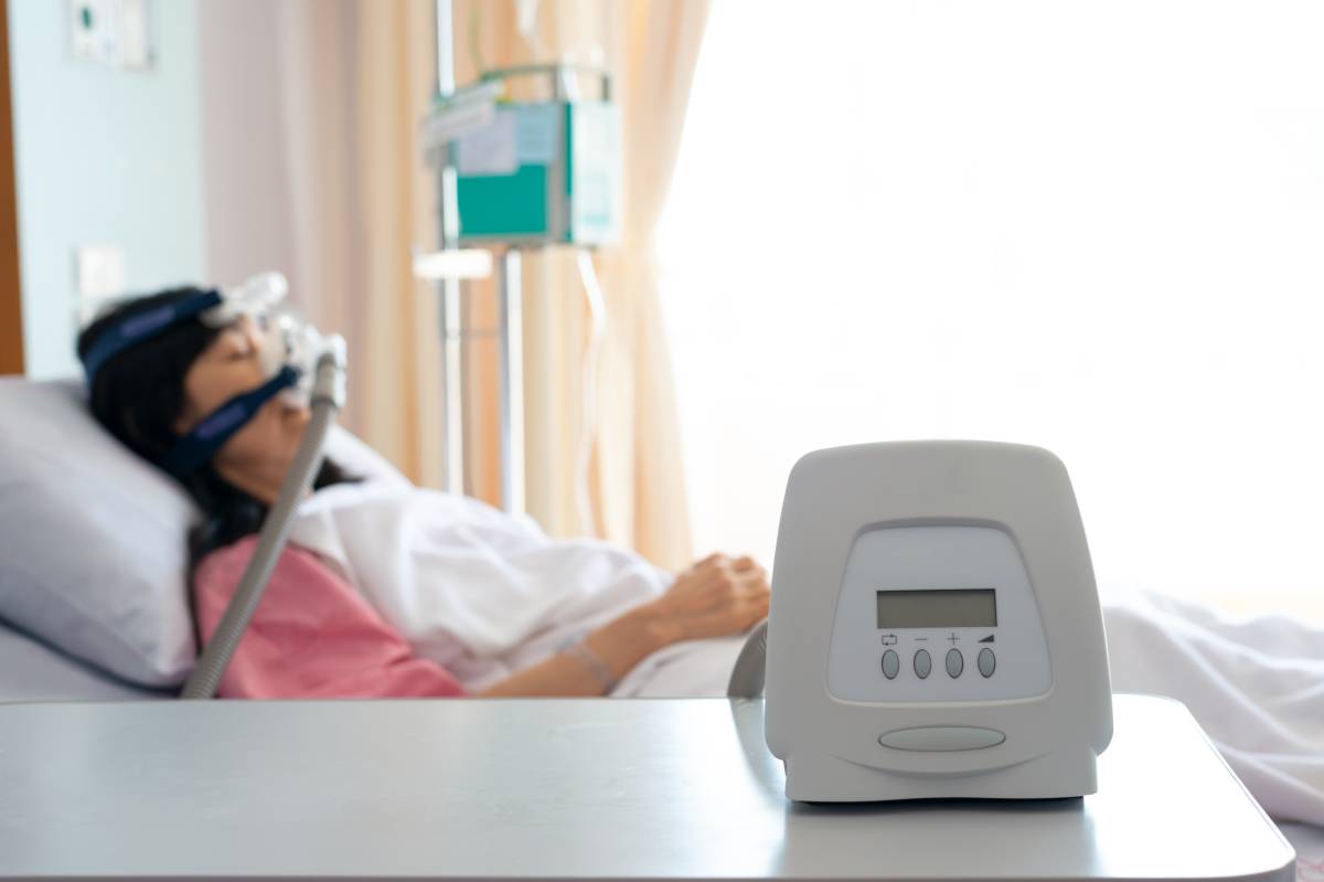
Appropriate treatments for sleep apnea can vary based on the severity of its presentation. For some patients, certain behavioral measures can result in a significant reduction in OSA symptoms. For instance, because obesity is a common risk factor for OSA and because weight-loss interventions have been associated with improvements in the severity of OSA, the American Thoracic Society recommends in their guidelines that clinicians incorporate weight management strategies into their treatment of overweight or obese adults with OSA.2 Independently of weight loss, exercise has also been associated with decreased severity in mild to moderate OSA,3  as well as abstaining from alcohol and restricting sleep to a side or prone position.1,4 For symptomatic OSA of any severity, positive airway pressure (PAP) devices are a primary therapy; PAP devices provide positive pressure to the airway through a mask to patients during sleep to keep the airway open and have been shown to reduce comorbidities such as high blood pressure in addition to alleviating the symptoms.5,6 Bariatric surgery to modify the upper airway is often recommended for symptomatic patients unable to tolerate PAP therapy, with the most extensively studied procedure being uvulopalatopharyngoplasty, a process involving the resection of the uvula and part of the soft palate.1 It is notable that more invasive treatments for OSA, such as surgical interventions, have been postponed or forgone for many patients during the COVID-19 pandemic.7 

Finally, hypoglossal nerve stimulation (HNS) therapy has recently been proposed as a potential treatment for OSA that could address the airway collapsibility associated with the disease without altering upper airway anatomy. This therapy, which involves a single surgical procedure to implant the device, has the potential to provide multilevel upper airway improvement and has shown early success within limited subsets of the OSA population.8 Another novel device designed to alleviate the symptoms of OSA was approved earlier this year by the U.S. Food and Drug Administration for marketing: the eXciteOSA device, designed for “tongue strengthening.” This device, used while the patient is awake, contains four electrodes which give muscle stimulation in a series of electrical pulses and is meant to increase the muscle tone of the tongue, which could actively work to help keep the airway open during sleep. While this device has only been used to aid those with mild sleep apnea and has not yet been proven to alleviate symptoms other than snoring, it is still an exciting development for a disorder for which novel treatments are sorely needed.9 

References 

(1)  Gottlieb, D. J.; Punjabi, N. M. Diagnosis and Management of Obstructive Sleep Apnea: A Review. JAMA 2020323 (14), 1389. https://doi.org/10.1001/jama.2020.3514

(2)  Hudgel, D. W.; Patel, S. R.; Ahasic, A. M.; Bartlett, S. J.; Bessesen, D. H.; Coaker, M. A.; Fiander, P. M.; Grunstein, R. R.; Gurubhagavatula, I.; Kapur, V. K.; Lettieri, C. J.; Naughton, M. T.; Owens, R. L.; Pepin, J.-L. D.; Tuomilehto, H.; Wilson, K. C. The Role of Weight Management in the Treatment of Adult Obstructive Sleep Apnea. An Official American Thoracic Society Clinical Practice Guideline. Am J Respir Crit Care Med 2018198 (6), e70–e87. https://doi.org/10.1164/rccm.201807-1326ST

(3)  Lee-Iannotti, J. K.; Parish, J. M. Exercise as a Treatment for Sleep Apnea. Journal of Clinical Sleep Medicine 202016 (7), 1005–1006. https://doi.org/10.5664/jcsm.8582

(4)  Srijithesh, P. R.; Aghoram, R.; Goel, A.; Dhanya, J. Positional Therapy for Obstructive Sleep Apnoea. Cochrane Database of Systematic Reviews 2019. https://doi.org/10.1002/14651858.CD010990.pub2

(5)  Weaver, T. E.; Maislin, G.; Dinges, D. F.; Bloxham, T.; George, C. F. P.; Greenberg, H.; Kader, G.; Mahowald, M.; Younger, J.; Pack, A. I. Relationship Between Hours of CPAP Use and Achieving Normal Levels of Sleepiness and Daily Functioning. Sleep 200730 (6), 711–719. https://doi.org/10.1093/sleep/30.6.711

(6)  Martínez-García, M.-A.; Capote, F.; Campos-Rodríguez, F.; Lloberes, P.; Díaz de Atauri, M. J.; Somoza, M.; Masa, J. F.; González, M.; Sacristán, L.; Barbé, F.; Durán-Cantolla, J.; Aizpuru, F.; Mañas, E.; Barreiro, B.; Mosteiro, M.; Cebrián, J. J.; de la Peña, M.; García-Río, F.; Maimó, A.; Zapater, J.; Hernández, C.; Grau SanMarti, N.; Montserrat, J. M. Effect of CPAP on Blood Pressure in Patients With Obstructive Sleep Apnea and Resistant Hypertension: The HIPARCO Randomized Clinical Trial. JAMA 2013310 (22), 2407. https://doi.org/10.1001/jama.2013.281250

(7)  Bastier, P.-L.; Aisenberg, N.; Durand, F.; Lestang, P.; Abedipour, D.; Gallet de Santerre, O.; Couloigner, V.; Bequignon, E. Treatment of Sleep Apnea by ENT Specialists during the COVID-19 Pandemic. European Annals of Otorhinolaryngology, Head and Neck Diseases 2020137 (4), 319–321. https://doi.org/10.1016/j.anorl.2020.05.001

(8)  Whelan, R.; Soose, R. J. Implantable Neurostimulation for Treatment of Sleep Apnea. Otolaryngologic Clinics of North America 202053 (3), 445–457. https://doi.org/10.1016/j.otc.2020.02.007

(9)  Mundell, E.; Reinberg, S. FDA Approves “Tongue Strengthening” Device for Certain Sleep Apnea Patients. Medical Press. February 8, 2021. 

Categories
Uncategorized

Lidocaine: Overview and Uses

Lidocaine is an intermediate-acting local anesthetic commonly used in surgical, obstetrical, and diagnostic procedure.1 This tertiary amine was first synthesized in the 1940s and quickly replaced several anesthetics in use at the time as a safer alternative.2 It is commonly administered with epinephrine to extend its duration of action and help prevent bleeding.3  

Lidocaine induces numbing by diffusing through neural sheaths and localizing to sodium channels, thus blocking the influx of sodium ions and preventing nerve depolarization.4 It has been found to have reduced efficacy in inflammatory conditions, which may be caused by an increase in blood flow or more acidic conditions that decrease the amount of un-ionized lidocaine molecules (the charged form is much less effective at diffusing through the cell membrane and subsequently acting on sodium channels).3  

It is most commonly used as a local numbing agent. In dental applications, a dilute solution is injected as close to the target nerve as possible. It is also commonly used to relieve the pain of post-herpetic neuralgia, the pain that can last months after a shingles infection, in the form of a transdermal patch.5 These patches usually consist of 5% lidocaine and can be used to treat pain from other sources, like post-operative nerve pain and compressed nerves. Traditionally, it has also been known to treat ventricular arrythmias, particularly ventricular tachycardia and multiple ventricular extrasystoles.6 However, more recent research suggests that, contrary to what was once believed, administering lidocaine prophylactically has a negligible effect on people who have experienced a myocardial infarction.7 

Though rare, adverse side effects of lidocaine have been observed. Most side effects are caused by toxic levels of lidocaine concentration in the plasma.This local anesthetic reaches toxic concentrations most easily when administered into the intercostal space, as it reaches the intravascular compartment most rapidly this way (as opposed to administration through the caudal, epidural, and subcutaneous spaces).More so than other anesthetics, lidocaine is associated with transient radicular irritation syndrome,8 which is lower extremity pain experienced after receiving spinal anesthesia.9 The drug is metabolized in the liver by the cytochrome CYP3A4, so any drugs that are also ligands of this can cause elevated lidocaine serum levels and may cause toxicity.10 

It has several contraindications. There are several cases in the literature of lidocaine-induced methemoglobinemia,11 a condition in which the iron in hemoglobin is stabilized in the ferric (Fe3+) form and cannot bind oxygen, which leads to tissue hypoxia.12 At high concentrations, it and other oxidizing drugs can convert hemoglobin to methemoglobin. Becker et al. stress the importance of hemodynamic monitoring when administering lidocaine with epinephrine or other vasoconstrictors, as, even in small doses, they can cause significant cardiovascular effects.3 

Foo et al. recently published a guide to using it intravenously in Anaesthesia, in which they recommend that infusions be given only in settings with continuous monitoring and after approval by the hospital.13 Lidocaine, the authors conclude, is a reliable and safe anesthetic that should be used by medical practitioners under the right conditions whenever it is needed. 

References 

1. “Lidocaine (Local) Monograph for Professionals.” Drugs.com.  

2. Calatayud, J., and González, A. “History of the Development and Evolution of Local Anesthesia Since the Coca Leaf.” Anesthesiology, vol. 98, no. 6, 2003, pp. 1503–1508., doi:10.1097/00000542-200306000-00031.  

3. Beecham GB, Bansal P, Nessel TA, et al. Lidocaine. [Updated 2021 Jan 26]. In: StatPearls [Internet]. Treasure Island (FL): StatPearls Publishing; 2021 Jan-. Available from: https://www.ncbi.nlm.nih.gov/books/NBK539881 

4. Lidocaine: A Common Local Anesthetic. The University of Reading. 

5. “Lidocaine Transdermal Patch: MedlinePlus Drug Information.” MedlinePlus, U.S. National Library of Medicine, medlineplus.gov/druginfo/meds/a603026.html.  

6. Gianelly, R., et al. “Effect of Lidocaine on Ventricular Arrhythmias in Patients with Coronary Heart Disease.” New England Journal of Medicine, vol. 277, no. 23, 1967, pp. 1215–1219., doi:10.1056/nejm196712072772301.  

7. Martí-Carvajal, A., et al. “Prophylactic Lidocaine for Myocardial Infarction.” Cochrane Database of Systematic Reviews, 2015, doi:10.1002/14651858.cd008553.pub2.  

8. Zaric, D, et al. “Transient Neurologic Symptoms (TNS) Following Spinal Anaesthesia with Lidocaine versus Other Local Anaesthetics.” Cochrane Database of Systematic Reviews, 2003, doi:10.1002/14651858.cd003006.  

9. Lambert, D. “Transient Radicular Irritation Remains a Danger.” Anesthesia Patient Safety Foundation, 20 Oct. 2019, www.apsf.org/article/transient-radicular-irritation-remains-a-danger/

10. Fang, P., et al. “Functional Assessment of CYP3A4 Allelic Variants on Lidocaine Metabolism in Vitro.” Drug Design, Development and Therapy, Volume 11, 2017, pp. 3503–3510., doi:10.2147/dddt.s152366.  

11. Chowdhary, S., et al. “Risk of Topical Anesthetic–Induced Methemoglobinemia.” JAMA Internal Medicine, vol. 173, no. 9, 2013, p. 771., doi:10.1001/jamainternmed.2013.75.  

12. Barash, M., et al. “Lidocaine-Induced Methemoglobinemia: A Clinical Reminder.” The Journal of the American Osteopathic Association, 2015, doi:10.7556/jaoa.2015.020.  

13. Foo, I., et al. “The Use of Intravenous Lidocaine for Postoperative Pain and Recovery: International Consensus Statement on Efficacy and Safety.” Anaesthesia, vol. 76, no. 2, 2020, pp. 238–250., doi:10.1111/anae.15270.