Categories
Uncategorized

Uses of Cameras in Surgery 

Minimally invasive surgery using a camera has arguably been the most important surgical advancement in the last three decades. It revolutionized surgical practice with well-demonstrated advantages over conventional open surgery, including decreased surgical trauma and incision-related complications such as surgical site infections, postoperative discomfort, and hernia, as well as a shorter hospital stay and better aesthetic result.4 Cameras allow surgeons to visualize and navigate the body during procedures without creating a large incision. Several different types of cameras are used in surgery, each with its unique features and advantages that have contributed to patient outcomes. 

Laparoscopic surgery uses cutting-edge technology to reduce tissue damage in which thin tubes called trocars are inserted in small “ports.” 2 A small camera (typically a laparoscope or endoscope) is then inserted into the trocars to view the procedures as a magnified image is projected to video monitors in the operating room.2 Depending on the operation, specialized equipment can also be introduced through the trocars. Not only do these techniques often provide the same results as traditional “open” surgery (which sometimes requires a big incision), but minimally invasive surgery (using tiny incisions) with cameras may also offer substantial advantages: (I) a quicker recovery; (II) a shorter length of hospitalization; (III) less scarring and tissue damage; and (IV) less discomfort.2 While laparoscopic surgical procedures has several advantages over traditional open surgery, acquiring the necessary skills, such as depth perception and video-hand-eye coordination, to move instruments within the operative field safely and effectively can be challenging.1 

Robotic surgery, which also uses cameras, has emerged as a new technique that is overcoming some difficulties of the standard laparoscopic approach in the field of hepatic, pancreatic, and esophageal surgery. It offers magnified three-dimensional optics, surgeon-controlled camera vision, working arms allowing very stable retraction, and unmatched ergonomics of instrument motion, with significantly less fatigue for the surgeon. 3 One of the major concerns regarding robotic technology is the high cost of equipment purchase and maintenance.3 Robotic surgery is more costly than laparoscopic or open surgery for various reasons, including equipment, higher operating time, and replacing materials as they wear out. Furthermore, the da Vinci surgical system is the only surgical robot in use.3 A lack of competition may be one of the factors keeping prices stable and high today.3 

With hundreds of millions of minimally invasive surgeries procedures performed globally, fiber optic cameras and robots have become indispensable tools and have revolutionized the field of surgery and surgeons’ capacity to capture information during surgery. 5 They have also helped to improve patient outcomes by reducing the risk of complications and the need for follow-up surgeries. However, it is important to note that using cameras in surgery does not eliminate the need for skilled surgeons. Cameras can only provide the surgeon with a visual aid; the surgeon’s skill and judgment ultimately determine the procedure’s success. 

References 

  1. Alaker, M., Wynn, G. R., & Arulampalam, T. (2016). Virtual reality training in laparoscopic surgery: A systematic review & meta-analysis. International journal of surgery (London, England), 29, 85–94. https://doi.org/10.1016/j.ijsu.2016.03.034 
  1. Baltayiannis, N., Michail, C., Lazaridis, G., Anagnostopoulos, D., Baka, S., Mpoukovinas, I., Karavasilis, V., Lampaki, S., Papaiwannou, A., Karavergou, A., Kioumis, I., Pitsiou, G., Katsikogiannis, N., Tsakiridis, K., Rapti, A., Trakada, G., Zissimopoulos, A., Zarogoulidis, K., & Zarogoulidis, P. (2015). Minimally invasive procedures. Annals of translational medicine, 3(4), 55. https://doi.org/10.3978/j.issn.2305-5839.2015.03.24 
  1. Biffi, R., Luca, F., Bianchi, P. P., Cenciarelli, S., Petz, W., Monsellato, I., Valvo, M., Cossu, M. L., Ghezzi, T. L., & Shmaissany, K. (2016). Dealing with robot-assisted surgery for rectal cancer: Current status and perspectives. World journal of gastroenterology, 22(2), 546–556. https://doi.org/10.3748/wjg.v22.i2.546 
  1. Bouquet de Joliniere, J., Librino, A., Dubuisson, J. B., Khomsi, F., Ben Ali, N., Fadhlaoui, A., Ayoubi, J. M., & Feki, A. (2016). Robotic Surgery in Gynecology. Frontiers in surgery, 3, 26. https://doi.org/10.3389/fsurg.2016.00026 
  1. Mascagni, P., Alapatt, D., Sestini, L., Altieri, M. S., Madani, A., Watanabe, Y., Alseidi, A., Redan, J. A., Alfieri, S., Costamagna, G., Boškoski, I., Padoy, N., & Hashimoto, D. A. (2022). Computer vision in surgery: from potential to clinical value. NPJ digital medicine, 5(1), 163. https://doi.org/10.1038/s41746-022-00707-5 
  1. Scognamiglio, P., Stüben, B. O., Heumann, A., Li, J., Izbicki, J. R., Perez, D., & Reeh, M. (2021). Advanced Robotic Surgery: Liver, Pancreas, and Esophagus – The State of the Art?. Visceral medicine, 37(6), 505–510. https://doi.org/10.1159/000519753 
Categories
Uncategorized

Post-Anesthesia Induction Hypotension 

Hypotension, also known as low blood pressure, can occur during or after anesthesia and must be watched closely by anesthesia providers. The post-anesthesia induction period may see increased risk for hypotension 1. Post-anesthesia induction hypotension (PAIH) can significantly impact surgical outcomes and remains one of the factors most closely associated with anesthesia-related morbidity 2. It is therefore critical to understand, predict and treat it as best as possible.  

Hypotension is roughly defined as a > 30% decrease in mean arterial pressure as compared to the first measurement in the operating theater prior to general anesthesia induction 3.  

In addition to heightened morbidity postoperatively, PAIH is associated with increased risk of renal injury and postoperative intensive care admission. It is also significantly linked to postoperative myocardial injury 2. Research has identified several risk factors: age, hypertension, diabetes, and being male. 

A recent multicenter observational study assessed the data of subjects receiving general anesthesia with propofol and sufentanil, demonstrating that that the likelihood of PAIH increased with age 1. Another study found that being over 30 years of age in particular was linked to PAIH 2. The degree of hypertension at time of arrival to the operating theater has also been found to be associated with PAIH 3, in addition to the presence of diabetes 3 and being male 1.   

Furthermore, PAIH has been clearly linked to the physical well-being of patients. One study found that it was linked to American Society of Anesthesiologists (ASA) physical status (PS) class IV patients, i.e. patients “with severe systemic disease that is a constant threat to life” 1,4. A more recent study found that patients with an ASA PS class II and above were more likely to experience PAIH 2.  

Different research has pointed to different links to the type of anesthesia used and the mode of delivery. First, early intraoperative hypotension in particular has been associated with neuraxial anesthesia 1. Second, although one study found that the type of volatile anesthetic was not linked to the occurrence of PAIH 3, another found that the administration of propofol and thiopental contributed to a greater incidence of PAIH 2. In addition, the type of surgery is also a relevant factor: orthopedic surgery in particular is associated with a greater incidence of PAIH 2.  

It remains unknown whether interventions to improve or maintain blood pressure would improve outcomes in patients with various risk factors. However, most clinicians err on the side of caution and try to avoid hypotension all together 6. A number of interventions exist to correct hypotension to this end, the overall efficacy of which exceed 94% 3. Bolus fluids are the most frequently used intervention, with an established effectiveness of 96% 3.  

Naturally, however, any factor linked to PAIH should be avoided when possible in order to minimize risk proactively. As such, one study suggests that alternatives to propofol anesthetic induction (such as etomidate) should be used in patients with an ASA PS of 3 or above and over 50 years of age 7

References 

1. Südfeld, S. et al. Post-induction hypotension and early intraoperative hypotension associated with general anaesthesia. Br. J. Anaesth. (2017). doi:10.1093/bja/aex127 

2. Nega, M. H., Ahmed, S. A., Tawuye, H. Y. & Mustofa, S. Y. Incidence and factors associated with post-induction hypotension among adult surgical patients: Prospective follow-up study. Int. J. Surg. Open 49, 100565 (2022). doi: 10.1016/j.amsu.2022.103321. 

3. Jor, O. et al. Hypotension after induction of general anesthesia: occurrence, risk factors, and therapy. A prospective multicentre observational study. J. Anesth. (2018). doi:10.1007/s00540-018-2532-6 

4. ​ASA Physical Status Classification System | American Society of Anesthesiologists (ASA). Available at: https://www.asahq.org/standards-and-guidelines/asa-physical-status-classification-system. (Accessed: 8th December 2022) 

5. Saugel, B. et al. Mechanisms contributing to hypotension after anesthetic induction with sufentanil, propofol, and rocuronium: a prospective observational study. J. Clin. Monit. Comput. (2022). doi:10.1007/s10877-021-00653-9 

6. Wong, G. T. C. & Irwin, M. G. Post-induction hypotension: a fluid relationship? Anaesthesia (2021). doi:10.1111/anae.15065 

7. Reich, D. L. et al. Predictors of hypotension after induction of general anesthesia. Anesth. Analg. (2005). doi:10.1213/01.ANE.0000175214.38450.91 

Categories
Uncategorized

Health Misinformation on Social Media

Access to accurate, up-to-date health information has been critical to keeping individuals and communities safe during the COVID-19 pandemic. As research around the virus evolved rapidly, social media became one site where health misinformation was widely disseminated—both intentionally and unintentionally (1). In response, U.S. Surgeon General Vivek Murthy has named Health Misinformation as one of his office’s top priorities (4). In an advisory titled “Confronting Health Misinformation,” Murthy stated that “Misinformation has caused confusion and led people to decline COVID-19 vaccines, reject public health measures such as masking and physical distancing, and use unproven treatments” (4). Within this landscape of widespread health misinformation on social media, it is critical that individuals, health professionals, companies, and governments take action and respond to this public health crisis. 

Health misinformation has been defined by researchers as health-related claims that are false or misleading according to current scientific consensus (3). Indeed, the spread of these false claims through social media is nothing new. A literature review from 2021 found that health misinformation was most common on topics of smoking and drugs, with the prevalence of health misinformation reaching 87% of posts in one category – Twitter posts about drugs – according to one study (5). Misinformation about vaccines had the second highest prevalence, reaching 43%, followed by diseases and pandemics at 40% and pro-eating disorder arguments at 36% (5). Amongst the platforms surveyed, Twitter had the highest rate of health misinformation (5). Some of these claims originated from non-medical professionals who are actively seeking to spread misinformation. However, many are simply the result of civilians reacting with confusion and fear. 

In order to remedy the surplus of health misinformation that circulates on social media, additional research and new initiatives are necessary at both local and large-scale levels. For instance, more extensive research needs to be conducted on understudied platforms such as Reddit or WeChat and non-textual content such as videos, images, and memes (3). Cross-disciplinary research is also necessary to understand psychological factors involved, considering that health topics can be intermingled with complex emotions (3). Finally, effective responses to health misinformation need to be developed. Simply refuting false claims may be ineffective in many situations (3). Proactive strategies, such as priming users with accurate information and educating individuals about identifying reliable sources, need to be balanced with reactive strategies. 

Individuals, healthcare professionals, and technology companies can all play a role in combating health misinformation on social media. Health professionals have the power to proactively engage with their social media audiences and provide factual information to patients and followers (2). In addition, technology platforms are being called to develop more effective strategies for monitoring content (1). Recently, Global Head of YouTube Health, Dr. Garth Graham, announced that the video platform will be incorporating health information panels to highlight authoritative sources and health content shelves that display reliable videos when users search for health-related topics (1). While it remains to be seen whether these strategies will be effective, individuals can take action in their online and in-person communities by vetting the health information they are presented on social media. 

References 

  1. Balsubramanian, Sai. “Health Misinformation Is A Pandemic, and Social Media Is Desperately Trying To Navigate it.” Forbes, 30 Oct 2022, www.forbes.com/sites/saibala/2022/10/30/health-misinformation-is-a-pandemic-and-social-media-is-desperately-trying-to-navigate-it/ 
  1. Bautista, John Robert et al. “Healthcare professionals’ acts of correcting health misinformation on social media.” International Journal of Medical Informatics, Vol. 148, April 2021, doi: 10.1016/j.ijmedinf.2021.104375 
  1. Chou, Wen-Ying et al. “Where We Go From Here: Health Misinformation on Social Media.” American Journal of Public Health, Vol. 110, No. S3, 2020, pp. S273-AS275, doi: 10.2105/AJPH.2020.305905 
  1. Murthy, Vivek. “Confronting Health Misinformation: The U.S. Surgeon General’s Advisory on Building a Healthy Information Environment.” Office of the U.S. Surgeon General, U.S. Department of Health and Human Services, www.hhs.gov/surgeongeneral/priorities/health-misinformation/index.html 
  1. Suarez-Lledo, Victor and Javier Alvarez-Galvez. “Prevalence of Health Misinformation on Social Media: Systematic Review.” Journal of Medical Internet Research, Vol 23, No. 1, Jan 20 2021, doi: 10.2196/17187
Categories
Uncategorized

Long Covid in Vaccinated vs. Unvaccinated Populations 

For most people, mild or moderate COVID-19 lasts for about two weeks. In others, however, health problems linger even after they are no longer testing positive for the illness; the long-term effects of coronavirus can persist for months or even years (Johns Hopkins Medicine, 2022). The World Health Organization describes post-COVID-19 condition, known colloquially as “long COVID,” as symptoms that persist or return “3 months from the onset of COVID-19… last for at least 2 months and cannot be explained by an alternative diagnosis” (WHO, 2021). Such symptoms can include fatigue, cognitive dysfunction (problems with thinking and memory), and shortness of breath, among others (WHO, 2021). They may be new following initial recovery from an acute COVID-19 episode, or they may persist from the initial illness; symptoms can also fluctuate or relapse over time. While getting vaccinated for COVID-19 does lower the risk of COVID infection, research concerning long COVID in vaccinated versus unvaccinated populations is ongoing. 

A recent study published in JAMA found that “among health care workers with SARS-CoV-2 infections not requiring hospitalization, 2 or 3 doses of vaccine, compared with no vaccination, were associated with lower long COVID prevalence” (Azzolini et al., 2022). Researchers from the Humanitas Research Hospital in Milan, Italy, conducted an observational cohort study from March 2020 to April 2022 among individuals working in 9 Italian health care facilities. All health care workers underwent weekly (in COVID wards) or biweekly (in other wards) PCR tests for COVID infection and had received three doses of the Pfizer-BioNTech vaccine over the course of 2021 (Azzolini et al., 2022). Researchers defined long COVID as reporting “at least 1 SARS-CoV-2-related symptom with a duration of more than 4 weeks” (Azzolini et al., 2022). Out of 2,560 participants, 29% had COVID-19, of which 31% had long COVID. Researchers categorized participants who caught COVID-19 by whether they were vaccinated at the time of infection and then calculated rates of long COVID for each group. Notably, having received more vaccine doses was associated with lower prevalence of long COVID: 41.8% when unvaccinated, 30.0% when having received 1 dose, 17.4% with 2 doses, and 16.0% with 3 doses of the vaccine (Azzolini et al., 2022). 

Long COVID has proved quite difficult to study, in part because the array of symptoms makes it hard to define. Even determining how common it is has been challenging: while some studies have previously suggested that long COVID occurs in as many as 30% of individuals infected with the virus, other results show much lower prevalence (Yoo et al., 2022; Stephenson et al., 2021). For example, a November 2021 study of around 4.5 million people treated at US Department of Veterans Affairs Hospitals suggests that the number is “7% overall and lower than that for those who were not hospitalized” (Xie et al., 2021). To date, there have been more than 93 million COVID-19 infections in the US alone (NYT, 2022). If even a small percentage of those infections turn into long COVID, “that’s a staggeringly high number of people affected by a disease that remains mysterious” (Reardon, 2022). To that end, some researchers suggest that vaccination alone might not be the best way to reduce the risk of long-term effects of Covid. Since research on long COVID is evolving, other COVID mitigation strategies remain important to the health of individuals worldwide. 

References 

Al-Aly, Z., Bowe, B., & Xie, Y. (2022). Long COVID after breakthrough SARS-CoV-2 infection. Nature Medicine, 28(7), 1461–1467. https://doi.org/10.1038/s41591-022-01840-0 

Azzolini, E., Levi, R., Sarti, R., Pozzi, C., Mollura, M., Mantovani, A., & Rescigno, M. (2022). Association Between BNT162b2 Vaccination and Long COVID After Infections Not Requiring Hospitalization in Health Care Workers. JAMA, 328(7), 676–678. https://doi.org/10.1001/jama.2022.11691 

Long COVID: Long-Term Effects of COVID-19. (2022, June 14). Johns Hopkins Medicine. https://www.hopkinsmedicine.org/health/conditions-and-diseases/coronavirus/covid-long-haulers-long-term-effects-of-covid19 

Reardon, S. (2022). Long COVID risk falls only slightly after vaccination, huge study shows. Nature. https://doi.org/10.1038/d41586-022-01453-0 

Stephenson, T., Shafran, R., & Rojas, N. (2021, August 10). Long COVID – the physical and mental health of children and non-hospitalised young people 3 months after SARS-CoV-2 infection; a national matched cohort study (The CLoCk) Study. Research Square. https://doi.org/10.21203/rs.3.rs-798316/v1 

Times, T. N. Y. (2020, March 3). Coronavirus in the U.S.: Latest Map and Case Count. The New York Times. https://www.nytimes.com/interactive/2021/us/covid-cases.html 

WHO: Clinical Services and Systems, Communicable Diseases, Technical Advisory Group on SARS-CoV-2 Virus Evolution. (2021, October 6). A clinical case definition of post COVID-19 condition by a Delphi consensus. World Health Organization. https://www.who.int/publications-detail-redirect/WHO-2019-nCoV-Post_COVID-19_condition-Clinical_case_definition-2021.1 

Xie, Y., Bowe, B., & Al-Aly, Z. (2021). Burdens of post-acute sequelae of COVID-19 by severity of acute infection, demographics and health status. Nature Communications, 12(1), 6571. https://doi.org/10.1038/s41467-021-26513-3 

Yoo, S. M., Liu, T. C., Motwani, Y., Sim, M. S., Viswanathan, N., Samras, N., Hsu, F., & Wenger, N. S. (2022). Factors Associated with Post-Acute Sequelae of SARS-CoV-2 (PASC) After Diagnosis of Symptomatic COVID-19 in the Inpatient and Outpatient Setting in a Diverse Cohort. Journal of General Internal Medicine, 37(8), 1988–1995. https://doi.org/10.1007/s11606-022-07523-3 

Categories
Uncategorized

Post Dural Puncture Headache 

Post Dural Puncture Headache

Neuraxial anesthesia refers to the administration of local anesthetic in or around the central nervous system (CNS), which blocks sensation for a certain region of the body. This type of anesthesia is often used for procedures in the lower body, sometimes in combination with general anesthesia (1). One potential side effect of neuraxial anesthesia and some spinal procedures is post dural puncture headache, and though advancements in knowledge and technology have drastically reduced its incidence, this condition causes serious discomfort and can negatively impact patient recovery (1-4). 

To understand post dural puncture headache, it is necessary to understand the anatomy of the meninges in the spine. Within the spinal column, the spinal cord is covered by three membranes – “meninges” – which are the dura mater (outermost), arachnoid mater, and pia mater (innermost). Cerebrospinal fluid (CSF) circulates within the meninges, as well as certain parts of the brain (1-3). Epidural anesthesia, which is one of the major subcategories of neuraxial anesthesia, delivers anesthetic to the space outside of the dura mater. Spinal anesthesia, the other major subcategory, delivers anesthetic to the space between the arachnoid and pia mater (1,2). 

Procedures that puncture the meninges, such as the administration of spinal anesthesia, have been linked to severe headaches when the patient is in the upright position. Procedures in the epidural space may inadvertently puncture the dura and cause post dural puncture headache as well (1-5). Patients may also experience dizziness, nausea and vomiting, and auditory or visual disturbances (2,3,5). There are several related hypotheses as to what causes these symptoms. Meningeal puncture allows CSF to leak out faster than it can be replenished naturally. Headache may be caused by resulting intracranial hypotension, compensatory vasodilation of vessels in the CNS, and/or mechanical stimulation of pain-sensitive structures in the skull due to the changing environment (2-5). Regarding the fact that post dural puncture headache tends to be more severe when upright, this is thought to be because the decreased level of CSF is magnified by gravity pulling the remaining fluid down and away from the brain. 

Several risk factors for post dural puncture headache have been identified. Young adults, women, lower BMI individuals, and those who experience chronic headache are more likely to experience this side effect after a spinal procedure. A more experienced provider performing the procedure, proper technique as elucidated by research, and smaller needle size are associated with lower risk (2,3,5). Accurate needle positioning can be more challenging in patients with obesity due difficulty palpating bony landmarks, which can lead to accidental dural puncture, but otherwise, higher BMI is actually associated with lower risk – one hypothesis for this pattern is that higher BMI results in higher intra-abdominal pressure that helps to counteract CSF leakage (3). 

The incidence of post dural puncture headache at the advent of neuraxial anesthesia was extremely high. However, research has revealed ways in which spinal procedure techniques can be modified to improve outcomes. These include smaller needles, non-cutting needles, and provider skill in the form of first pass success (3,5). 

Treatment for this condition is still a topic of research; currently accepted approaches include inducing vasoconstriction (such as with caffeine) and placing a “blood patch” to generate a clot that blocks the puncture. Note, however, that post dural puncture headache does resolve on its own given time (2,4). 

References 

  1. Olawin AM, Das JM. Spinal Anesthesia. StatPearls [Internet]. 2021. https://www.ncbi.nlm.nih.gov/books/NBK537299/ 
  1. Turnbull DK, Shepherd DB. Post‐dural puncture headache: pathogenesis, prevention and treatment. British Journal of Anaesthesia. 2003; 91(5):718–729. DOI:10.1093/bja/aeg231 
  1. Harrington BE, Reina MA. “Postdural puncture headache.” NYSORA. (n.d.). https://www.nysora.com/topics/complications/postdural-puncture-headache/ 
  1. Committee on Obstetric Anesthesia. “Statement on Post-Dural Puncture Headache Management.” ASA. 2021. https://www.asahq.org/standards-and-guidelines/statement-on-post-dural-puncture-headache-management/ 
  1. Kim JE, Kim SH, Han RJW, et al. Postdural Puncture Headache Related to Procedure: Incidence and Risk Factors After Neuraxial Anesthesia and Spinal Procedures. Pain Medicine. 2021;22(6):1420-1425. DOI:10.1093/pm/pnaa437 
Categories
Uncategorized

Health System Responses to Data Breaches 

One of the fundamental beliefs within the American medical system is that patients have the right to privacy. This right is increasingly challenged by cyberattacks, and thus how the health system should respond to data breaches is a key area of work. According to the North American Association for Central Cancer Registries,  

“Confidentiality is the cancer registry’s responsibility to the patients whose data are in the database and is of paramount concern to all cancer registries. There may be no greater threat to the operation and maintenance of a cancer registry than an actual or perceived breach of confidentiality. In fact, an actual or perceived breach of confidentiality in one registry may threaten all registries.”1 

That is to say – to threaten this privacy is to threaten the practice of keeping patient records at all, which in turn threatens the practice of medicine as we know it. 

Although there are many measures of security to protect both the healthcare workplace and its respective databases, data breaches are an unfortunate eventuality. A data breach can occur for any number of reasons: an accidental violation of HIPAA protocol, for example, or a pre-planned attack by a hacker hoping to negotiate ransom. No matter the type of breach, it is critical that healthcare providers have both protection against breaches as well as a response protocol. 

According to the CDC, successful management of a data breach starts long before the incident even occurs. In other words, a pre-written detailed plan in the case of a data breach should be organized and shared amongst healthcare employees to ensure rapid response. Once such a plan has been drafted, agreed upon, and taught, “it is the program’s responsibility to execute its response plan.”2 Failure to do so increases the risk of violating legislative protocol,3 worsening the impact of the original breach, and enabling subsequent breaches. These in turn can cause the healthcare institution to lose credibility with patients and other healthcare providers, as well as cause harm to patients themselves 

One key part of said plan is a breach response team (BRT), or a group of people with the designated responsibility of investigating suspected data breaches in a health system. It is advisable that the members of such team have a background in computer science or information technology, which will allow them to troubleshoot each incident.2 Familiarity with each facility’s technology and security measures is also a prerequisite for being a member of the BRT. Duties of the BRT can include (but is not limited to) developing detection programs and methods for reporting breaches, responding to and tracking suspected breaches, evaluating response tactics, and notifying individuals whose privacy may have been affected by the data breach. However, it is not the job of the BRT alone to manage data breaches. The workplace as a whole must be well-educated and ready to respond in the case of a breach. If proper education is giving and non-compliance leads to a data breach, then that individual employee is responsible and can face both legal and corporate charges. Even an accidental breach may culminate in loss of employment and the potential for legal repercussions. 

Clearly, protection of private data is integral to the function and purpose of a healthcare facility. Therefore, responding to data breaches in a timely, effective, and appropriate manner is of utmost importance. 

References 

1 Standards for completeness, quality, analysis, and management of data, Volume III. NAACCR. (2019, September 12). Retrieved from https://www.naaccr.org/standards-for-completeness-quality-analysis-and-management-of-data/  

2 Centers for Disease Control and Prevention. (2021, January 20). Data breach response. Centers for Disease Control and Prevention. Retrieved from https://www.cdc.gov/cancer/npcr/tools/security/breach.htm  

3 (OCR), O. for C. R. (2021, June 28). Breach notification rule. HHS.gov. Retrieved from https://www.hhs.gov/hipaa/for-professionals/breach-notification/index.html 

Categories
Uncategorized

R-Ketamine vs. S-Ketamine

(R,S)-ketamine is a N-methyl-ᴅ-aspartate (NMDA) receptor antagonist and a commonly used anesthetic agent worldwide. In the late 1990s, studies and case reports began highlighting this drug’s rapid-acting and sustained antidepressant effects, a major discovery in the research of mood disorders [1]. (R,S)-ketamine is a mixture of the two enantiomers R- and S-ketamine, which predominantly differ in their binding properties [2]. S-ketamine has an approximately fourfold greater affinity for the phencyclidine site of the NMDA receptor than R-ketamine as well as strong anti-depressant effects but also strong psychomimetic side effects such as confusion, euphoria, perceptual difficulties, or mood elevation. On the other hand, R-ketamine is generally associated with a milder but longer-lasting antidepressant effect [2,3].

In 2017, a PET study on conscious monkeys found a reduction of dopamine D2/3 binding potential in the striatum following S-ketamine administration, but not R-ketamine [4]. In 2000, a research team in Japan used monkey brains and found [11C] raclopride could be used in PET to detect release of endogenous dopamine from presynaptic terminals [5]. Applying this finding, researchers at the Central Research Laboratory in Japan (2017) found marked radioactivity in the striatum of S-ketamine-treated animals, suggesting S-ketamine causes significant release of dopamine but prevents it from binding to its receptors [4]. The excessive dopamine may also be the cause of the psychosis and dissociation associated with chronic S-ketamine administration. In healthy subjects, an infusion of S-ketamine produced a dissociative state, changes in mood and sensory perception, difficulty in reality appraisal, and ego inflation [6]. Despite an increasing number of studies arguing in favor of ketamine’s role in treating depressive disorders, the drug’s abuse potential is its greatest limitation. The conditioned place preference test (CPP) is a widely used behavioral model designed to assess a drug’s rewarding, aversive, or addicting effects [7].  A 2015 preclinical study found ketamine (the racemic combination) increased scores on the CPP test, suggesting ketamine itself is rewarding [8]. Another preclinical study used the same assessment and found similar increases after S-ketamine administration, but not R-ketamine, indicating potential abuse liability of S-ketamine in particular [9].

Parvalbumin (PV) positive cells are GABAergic interneurons that rely on Ca2+ binding for proper functioning [10]. A reduction in PV-neurotransmission is associated with neuropsychiatric disorders such as Alzheimer’s Disease, autism spectrum disorder, schizophrenia, and substance use disorder. In 2016, a research team in Japan used PV-immunohistochemistry to assess the effect of intermittent ketamine administration. They observed a significant decrease in PV-immunoreactivity in the prelimbic and infralimbic areas of the prefrontal cortex, as well as the CA1, CA3, and dentate gyrus of the hippocampus after intermittent administrations of S-ketamine, but not R-ketamine [11]. These results correlate with earlier findings using the same methods but with single-dose administrations of the respective drugs [8].  These studies suggest S-ketamine plays a role in the loss of PV-positive cells, which is associated with psychiatric presentations [11].

S-ketamine is metabolized to its major metabolite, S-norketamine, by cytochrome P450 enzymes. Like S-ketamine, S-norketamine induces strong antidepressant effects in murine models of depression. S-norketamine significantly attenuated the reduced dendritic spine density in the prelimbic area, the CA3, and the dentate gyrus in mice exposed to chronic social defeat stress. In the same regions, S-norketamine also improved reduced levels of BDNF protein, a marker of neuroplasticity [12]. Unlike S-ketamine, its metabolite does not show psychomimetic effects such as increased locomotion, hyperactivity, or increased scores on the conditioned place preference test. S-norketamine also had no effect on the proportion of PV-positive cells in the prefrontal cortex [9,12]. These results suggest that S-norketamine may be a more promising therapeutic approach, if it can be successfully stabilized and administered.

Although the US and Europe approved an S-ketamine based nasal spray for treatment-resistant depression, several concerns have been raised, including its safety for pediatric patients and its numerous side effects [13,14]. A recent pilot study demonstrated R-ketamine produced sustained antidepressant effects without side effects such as dissociation [9,15]. With ketamine’s different enantiomers and metabolites, more research on their effects and applications is warranted. 

References

  1. Abdallah, C. G., Sanacora, G., Duman, R. S., & Krystal, J. H. (2018). The Neurobiology of Depression, Ketamine and Rapid-acting Antidepressants: Is it Glutamate Inhibition or Activation? Pharmacology & Therapeutics190, 148–158. https://doi.org/10.1016/j.pharmthera.2018.05.010
  2. Paul, R., Schaaff, N., Padberg, F., Möller, H.-J., & Frodl, T. (2009). Comparison of Racemic Ketamine and S-ketamine in Treatment-resistant Major Depression: Report of Two Cases. The World Journal of Biological Psychiatry: The Official Journal of the World Federation of Societies of Biological Psychiatry10(3), 241–244. https://doi.org/10.1080/15622970701714370
  3. Zhang, J., Li, S., & Hashimoto, K. (2014). R(−)Ketamine shows Greater Potency and Longer Lasting Antidepressant Effects than S (+)Ketamine. Pharmacology Biochemistry and Behavior116, 137–141. https://doi.org/10.1016/j.pbb.2013.11.033
  4. Hashimoto, K., Kakiuchi, T., Ohba, H., Nishiyama, S., & Tsukada, H. (2017). Reduction of Dopamine D2/3 Receptor Binding in the Striatum after a Single Administration of Esketamine, but not R-Ketamine: A PET study in Conscious Monkeys. European Archives of Psychiatry and Clinical Neuroscience267(2), 173–176. https://doi.org/10.1007/s00406-016-0692-7
  5. Tsukada, H., Harada, N., Nishiyama, S., Ohba, H., & Kakiuchi, T. (2000). Cholinergic Neuronal Modulation Alters Dopamine D2 Receptor Availability in vivo by Regulating Receptor Affinity Induced by Facilitated Synaptic Dopamine Turnover: Positron Emission Tomography Studies with Micro-dialysis in the Conscious Monkey Brain. Journal of Neuroscience20(18), 7067–7073. https://doi.org/10.1523/JNEUROSCI.20-18-07067.2000
  6. Vollenweider, F. X., Leenders, K. L., Øye, I., Hell, D., & Angst, J. (1997). Differential Psychopathology and Patterns of Cerebral Glucose Utilization Produced by (S)- and (R)-ketamine in Healthy Volunteers using Positron Emission Tomography (PET). European Neuropsychopharmacology7(1), 25–38. https://doi.org/10.1016/S0924-977X(96)00042-9
  7. Prus, A. J., James, J. R., & Rosecrans, J. A. (2009). Conditioned Place Preference. In J. J. Buccafusco (Ed.), Methods of Behavior Analysis in Neuroscience (2nd ed.). CRC Press/Taylor & Francis. http://www.ncbi.nlm.nih.gov/books/NBK5229/
  8. Yang, C., Shirayama, Y., Zhang, J. -c, Ren, Q., Yao, W., Ma, M., Dong, C., & Hashimoto, K. (2015). R-Ketamine: A Rapid-onset and Sustained Antidepressant Without Psychotomimetic Side Effects. Translational Psychiatry5(9), e632–e632. https://doi.org/10.1038/tp.2015.13
  9. Hashimoto, K. (2020). Molecular Mechanisms of the Rapid-acting and Long-lasting Antidepressant Actions of (R)-ketamine. Biochemical Pharmacology177, 113935. https://doi.org/10.1016/j.bcp.2020.113935
  10. Nahar, L., Delacroix, B. M., & Nam, H. W. (2021). The Role of Parvalbumin Interneurons in Neurotransmitter Balance and Neurological Disease. Frontiers in Psychiatry12. https://www.frontiersin.org/article/10.3389/fpsyt.2021.679960
  11. Yang, C., Han, M., Zhang, J., Ren, Q., & Hashimoto, K. (2016). Loss of Parvalbumin-Immunoreactivity in Mouse Brain Regions After Repeated Intermittent Administration of Esketamine, but not R-ketamine. Psychiatry Research239, 281–283. https://doi.org/10.1016/j.psychres.2016.03.034
  12. Yang, C., Kobayashi, S., Nakao, K., Dong, C., Han, M., Qu, Y., Ren, Q., Zhang, J., Ma, M., Toki, H., Yamaguchi, J., Chaki, S., Shirayama, Y., Nakazawa, K., Manabe, T., & Hashimoto, K. (2018). AMPA Receptor Activation–Independent Antidepressant Actions of Ketamine Metabolite (S)-norketamine. Biological Psychiatry84(8), 591–600. https://doi.org/10.1016/j.biopsych.2018.05.007 
  13. Zimmermann, K. S., Richardson, R., & Baker, K. D. (2020). Esketamine as a Treatment for Pediatric Depression: Questions of Safety and Efficacy. The Lancet Psychiatry7(10), 827–829. https://doi.org/10.1016/S2215-0366(19)30521-8
  14. Turner, E. H. (2019). Esketamine for Treatment-resistant Depression: Seven Concerns about Efficacy and FDA Approval. The Lancet Psychiatry6(12), 977–979. https://doi.org/10.1016/S2215-0366(19)30394-3
  15. Leal, G. C., Bandeira, I. D., Correia-Melo, F. S., Telles, M., Mello, R. P., Vieira, F., Lima, C. S., Jesus-Nunes, A. P., Guerreiro-Costa, L. N. F., Marback, R. F., Caliman-Fontes, A. T., Marques, B. L. S., Bezerra, M. L. O., Dias-Neto, A. L., Silva, S. S., Sampaio, A. S., Sanacora, G., Turecki, G., Loo, C., … Quarantini, L. C. (2021). Intravenous Arketamine for Treatment-resistant Depression: Open-label Pilot Study. European Archives of Psychiatry and Clinical Neuroscience271(3), 577–582. https://doi.org/10.1007/s00406-020-01110-5
Categories
Uncategorized

Silent Hypoxia in COVID-19 

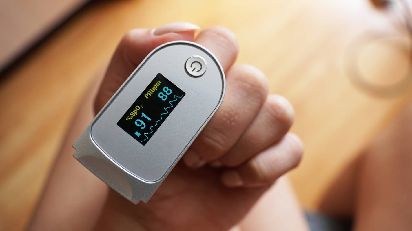
Silent hypoxia is characterized by a significantly reduced oxygen saturation level with no associated breathing difficulties, at least in its beginning stages [1]. Because patients do not present with trouble breathing until the condition has progressed significantly, it can be difficult to detect [1]. Studies have recently reported that 20 to 40% of COVID-19 patients may experience silent hypoxia [1]. By the time medical teams have detected silent hypoxia, the patient may already have progressed into moderate-to-severe levels of infection, worsening their long-term recovery rates [2]. Consequently, medical practitioners must familiarize themselves with the warning signs of silent hypoxia to detect and treat it early. 

There are many possible reasons why COVID-19 patients may develop silent hypoxia and, subsequently, experience a deterioration of their ability to breathe. Researchers have posited various hypotheses, from the virus’s impact on blood vessels contributing to impaired hypoxic vasoconstriction to  changes to one’s respiratory system, perhaps via the expression of angiotensin-converting enzyme-2 receptors in the carotid bodies and nasal mucosa [1][3]. Unfortunately, while there are many possible theories concerning the development of this condition, research has not yet isolated the principal answer, making treatment more difficult [1].  

While the underlying mechanisms guiding the development of silent hypoxia may remain uncertain, researchers have made progress in identifying certain predictors for the condition. In a retrospective cohort study, Alhusain and colleagues analyzed data from all of the COVID-19 patients who visited a hospital in Riyadh, Saudi Arabia over ten months [4]. Among the 195 patients who experienced silent hypoxia, the researchers identified no correlations between dyspnea and gender, age group, comorbidity, or body mass index [4]. This finding was partially consistent with another study by Okhuama et al., who found that age and diabetes were non-predictors of silent hypoxia in COVID-19 patients and suggested that more complex mechanisms were at play [5]. However, Alhusain et al. did find that fever, cough, and chronic cardiac disease were predictors of dyspnea, providing medical teams with some guidance in anticipating the condition [4]. 

Medical teams can increase their chances of successfully identifying silent hypoxia by monitoring certain elements of the patient’s profile. Given that silent hypoxia patients may suffer from levels of pneumonia that are disproportionate to their COVID symptoms, practitioners should closely monitor patients’ pulse oximetry and arterial blood gas levels [5]. Bluish coloration can also indicate that a COVID-19 patient has silent hypoxia [6]. Before getting to the hospital setting, patients can also engage in detection practices themselves by regularly checking their blood saturation levels using a pulse oximeter or a smartphone [2]. A drop in oxygen saturation levels below 95% warrants contacting a health provider [2]. 

Ultimately, silent hypoxia is a pressing condition that can increase rates of intubation, mechanical ventilation, and even death among COVID-19 patients [2]. While medical teams still await more information about silent hypoxia, heeding the aforementioned indications can help treat pandemic patients and, moreover, suppress the adverse impacts of this ongoing pandemic. 

References 

[1] A. Rahman et al., “Silent hypoxia in COVID-19: pathomechanism and possible management strategy,” Molecular Biology Reports, vol. 48, no. 4, p. 3863-3869, April 2021. [Online]. Available: https://doi.org/10.1007/s11033-021-06358-1

[2] J. Teo, “Early Detection of Silent Hypoxia in Covid-19 Pneumonia Using Smartphone Pulse Oximetry,” Journal of Medical Systems, vol. 44, no. 8, p. 1-2, June 2020. [Online]. Available: https://doi.org/10.1007/s10916-020-01587-6

[3] T. Fuehner et al., “Silent Hypoxia in COVID-19: A Case Series,” Respiration, vol. 101, p. 376-380, November 2021. [Online]. Available: https://doi.org/10.1159/000520083

[4] F. Alhusain et al., “Predictors and clinical outcomes of silent hypoxia in COVID-19 patients, a single-center retrospective cohort study,” Journal of Infection and Public Health, vol. 14, no. 11, p. 1595-1599, November 2021. [Online]. Available: https://doi.org/10.1016/j.jiph.2021.09.007

[5] A. Okuhama et al., “Clinical and radiological findings of silent hypoxia among COVID-19 patients,” Journal of Infection and Chemotherapy, vol. 27, no. 10, p. 1536-1538, October 2021. [Online]. Available: https://doi.org/10.1016/j.jiac.2021.07.002

[6] T. S. Simonson et al., “Silent hypoxaemia in COVID-19 patients,” The Journal of Physiology, vol. 599, no. 4, p. 1057-1065, December 2020. [Online]. Available: https://doi.org/10.1113/JP280769

Categories
Uncategorized

Anesthesia Considerations for Balloon Sinuplasty

In 2005, the FDA approved balloon sinus ostial dilation–also known as balloon sinuplasty, or BSD–for use in treating both recurrent acute and chronic rhinosinusitis [1]. This technique involves placing a balloon catheter into the sinus ostium; the resultant inflation dilates the sinus opening and subsequently relieves obstructions [1]. Since becoming FDA-approved, several studies have confirmed the safety of balloon sinuplasty procedures and have discovered additional benefits, including, but not limited to, avoidance of general anesthesia in certain cases [1]. 

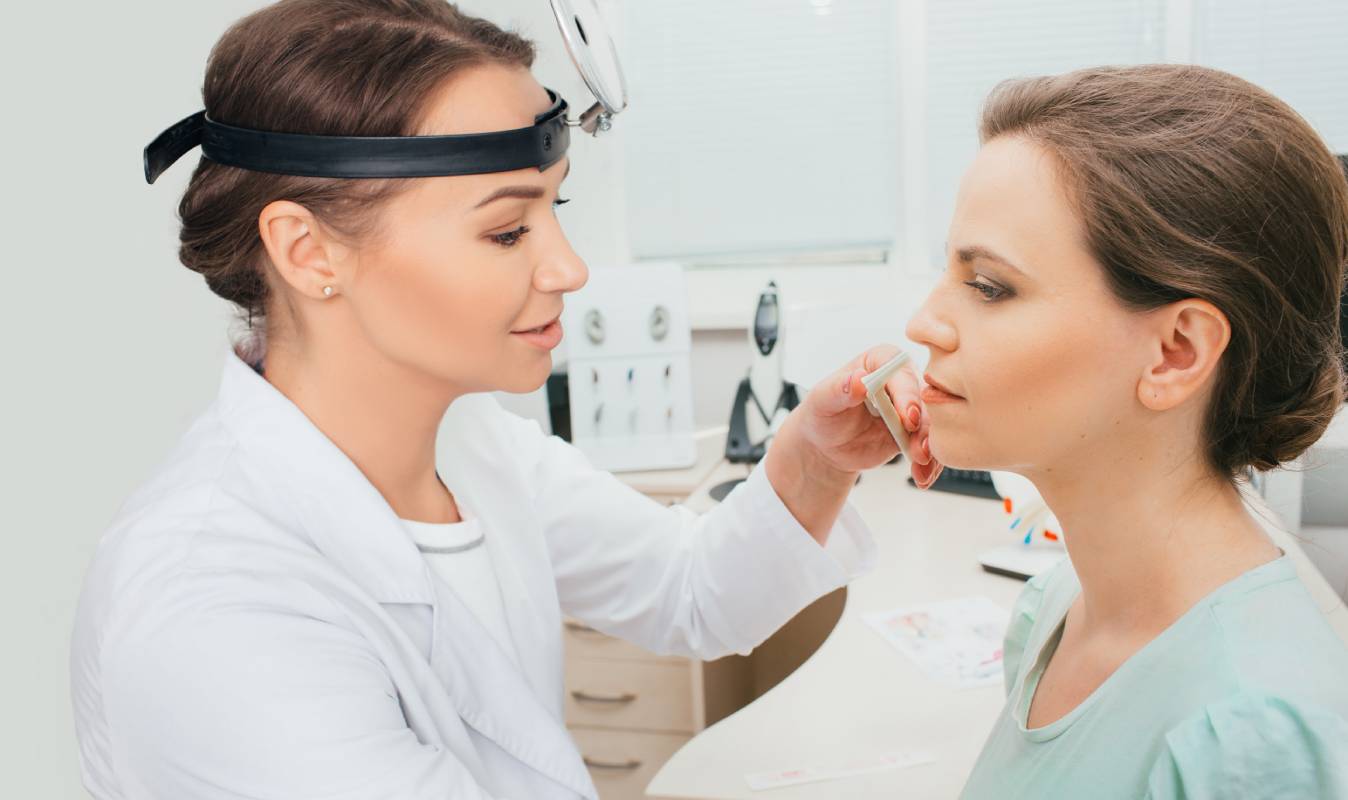
During balloon sinuplasty, a patient requires anesthesia [2]. When the procedure was first introduced, patients typically received general anesthesia by way of an endotracheal tube or a laryngeal mask airway [2]. However, since its introduction, sedation and local anesthesia have become more common, facilitating a transition to office-based procedures [2]. Compared with sinus surgery performed under general anesthesia, local anesthesia-based balloon sinuplasty has the advantages of greater convenience, quicker recovery, similar technical success, and lower costs [3]. 

In a recent study, Naik et al. compared local and general anesthesia [4]. They split their 50 subjects into two evenly sized groups: one that underwent BSP with general anesthesia, while the other received a combined nerve block and topical anesthesia regimen [4]. The researchers found that the local anesthesia group experienced a shorter time gap between when the patient entered the operating theater and when the surgical procedure began; they also recovered quicker [4]. However, this group reported feeling more intraoperative discomfort than the general anesthesia group [4]. Additionally, surgeons performing local anesthesia were less comfortable than those treating the general anesthesia patients [4]. Nevertheless, Naik and colleagues concluded that, for “less apprehensive and motivated cases,” anesthesia providers can opt for local anesthetics which, along with promoting quicker results, are also the more cost-effective option [4].

Although it is considered an alternative to endoscopic sinus surgery (ESS), BSD is more accurately described as another tool, device, or instrument available to physicians performing ESS [1]. Instead of placing a balloon in the sinuses, physicians performing traditional ESS place an endoscope or, in more difficult cases, more specialized instruments to clear drainage pathways and potentially straighten the septum. [5]. Despite being less invasive than ESS, BSD has significant similarities with ESS in terms of anesthetic techniques [6, 7]. Therefore, when considering best anesthetic practices to use in conjunction with BSD, guidelines regarding anesthesia administration during ESS may also be relevant for anesthesia providers performing general anesthesia-based BSD.

Accordingly, anesthesia providers should take steps to avoid or minimize surgical bleeding [8]. To minimize the occurrence of surgical bleeding–the most serious form of which is capillary bleeding–some research points toward avoiding volatile anesthetic agents, which cause vasodilation, where possible [8]. Additionally, maintaining anesthesia depth by providing patients with muscle relaxants and limiting the use of positive end-expiratory pressure can prevent higher intrathoracic pressure, which increases surgical bleeding from the head [8]. Maintaining normothermia is also important for minimizing bleeding, as is administering local anesthetics and vasoconstrictors [8].

Researchers are optimistic that, in the future, patients may require minimal to no anesthesia when undergoing balloon sinuplasty [2]. While anesthetics are still necessary, opting against general anesthesia when possible and, when it is not, minimizing surgical bleeding is important to the success of BSD-based surgeries.

References 

[1]  C. Cingi, N. Bayar Muluk, and J. T. Lee, “Current indications for balloon sinuplasty,” Current Opinion in Otolaryngology & Head and Neck Surgery, vol. 27, no. 1, p. 7-13, February 2019. [Online]. Available: https://doi.org/10.1097/MOO.0000000000000506

[2]  A. E. Stewart and W. C. Vaughan, “Balloon Sinuplasty Versus Surgical Management of Chronic Rhinosinusitis,” Current Allergy and Asthma Report, vol. 10, 1, p. 181-187, March 2010. [Online]. Available: https://doi.org/10.1007/s11882-010-0105-3

[3]  J. Gould et al., “In-Office, Multisinus Balloon Dilation: 1-Year Outcomes from a Prospective, Multicenter, Open Label Trial,” American Journal of Rhinology & Allergy, vol. 28, no. 2, p. 156-163, March-April 2014. [Online]. Available: https://doi.org/10.2500/ajra.2014.28.4043

[4]  S. S. Naik, C. Venkategowda, N. Reddy, and S. M. Naik, “Combined Nerve Block and Topical Anesthesia: An Effective Alternate to General Anesthesia in Hybrid Balloon Sinuplasty Procedures,” Journal of Research & Innovation in Anesthesia, vol. 5, no. 1, p. 6-9, January-June 2020. [Online]. Available: https://doi.org/10.5005/jp-journals-10049-0080

[5] “Balloon Sinuplasty v. Endoscopic Sinus Surgery Explained,” Kaplan Sinus Relief, Updated October 23, 2020. [Online]. Available: https://www.kaplansinusrelief.com/blog/balloon-sinuplasty-vs-endoscopic-sinus-surgery-explained/

[6]  A. Koskinen et al., “Comparison of intra-operative characteristics and early post-operative outcomes between endoscopic sinus surgery and balloon sinuplasty,” Acta Oto-Laryngologica, vol. 137, no. 2, p. 202-206, April 2018. [Online]. Available: https://doi.org/10.1080/00016489.2016.1227476

[7]  J. Flávio Nogueira Júnior, A. C. Stamm, and S. Pignatari, “Balloon sinuplasty, an initial assessment: 10 cases, results and follow-up,” Brazilian Journal of Otorhinolaryngology, vol. 76, no. 5, p. 588-595, September/October 2010. [Online]. Available: https://doi.org/10.1590/S1808-86942010000500009

[8]  P. Y. Tan and R. Poopalalingam, “Anaesthetic Concerns for Functional Endoscopic Sinus Surgery,” Proceedings of Singapore Healthcare, vol. 23, no. 3, p. 246-253, 2014. [Online]. Available: https://doi.org/10.1177/201010581402300310

Categories
Uncategorized

Anesthesiology vs. Pain Medicine

Historically, pain medicine was a subset of the general field of anesthesiology [1]. With the advent of nerve procedures, however, the two disciplines evolved to encompass increasingly separate, but nevertheless interrelated, functions [1]. These two fields have differing roles, educational requirements, and treatment options.

Pain medicine doctors, also referred to as pain management doctors, specialize in the diagnosis, management, and treatment of pain and painful disorders [2]. They assist patients with varying pain levels, ranging from acute to chronic [2]. Alternatively, anesthesiologists are experts in providing medical care for patients at every step of surgery [3]. Their major focus is sustaining life function during operations, which means that they are trained in pain management, but approach pain from a specific perspective [3].

There are two categorizations within the overall discipline of pain medicine: “medical pain” and interventional [1]. Medical pain is the broader category. Practitioners in this field can include family medicine doctors, internists, and psychiatrists [1]. Accordingly, they come from a diverse range of educational backgrounds–pain medicine doctors may have completed residencies in neurology, psychiatry, rehabilitation, or physical medicine, among other disciplines [2]. They primarily work with people who suffer from chronic ailments and, therefore, may require long-term medical treatment and, in some cases, opioids or other medications [1]. These doctors can prescribe a varied range of treatment options, either separately or in combination, to alleviate patients’ pain [4]. For instance, physicians may recommend interdisciplinary treatment plans that combine physical therapy, psychotherapy, and acupuncture [4].

While the distinction between anesthesiologists and the doctors providing care for pain relief as described above is evident, the boundary between pain medicine and anesthesiology is more blurred when considering interventional pain physicians. Interventional doctors treat their patients by way of more complicated pain management procedures and techniques [1]. They can administer spine injections, nerve blocks, and implantable devices [1]. As such, they are often trained in anesthesiology. In the United States, interventional pain management physicians must finish one year of internship, a residency in either anesthesiology, neurology, psychiatry or rehabilitative medicine, and a one-year-long fellowship in pain management [1].

Today, there remains some debate about the intersection between anesthesiology and pain management, most notably pertaining to the treatment of chronic pain. Anesthesiologists receive specialized training in acute pain, but they do not necessarily study chronic pain management [5]. As a result, some physicians have advocated for educational reform that would either incorporate chronic pain training into the anesthesiology curriculum or further distinguish chronic pain treatment from anesthesiology [5]. While it is unclear which path the medical community will take, the dual training that many interventional pain doctors receive in anesthesiology and pain management appears to be a step in the right direction.

Millions of patients experience pain every year [6]. Fortunately, anesthesiologists and pain management doctors can offer a diverse range of treatment options to their patients. By keeping in mind the respective strengths of these two classes of practitioners, patients will hopefully have a greater chance of receiving appropriate and effective pain relief.

References 

[1] “What Does a Pain Management Doctor Do?,” Integris Health, Updated September 21, 2020. [Online]. Available: https://integrisok.com/resources/on-your-health/2020/september/what-does-a-pain-management-doctor-do

[2] S. Lewis, “Pain Medicine Doctor: Your Pain Relief & Pain Management Specialist,” Healthgrades, Updated December 21, 2017. [Online]. Available: https://bit.ly/3oWLj2X

[3] S. Lewis, “Anesthesiologist: Your Surgical Anesthesia & Pain Management Specialist,” Healthgrades, Updated January 21, 2020. [Online]. Available: https://bit.ly/3oU5O08

[4] “What does a pain management doctor do?,” Drugs.com, Updated August 24, 2021. [Online]. Available: https://www.drugs.com/medical-answers/pain-management-doctor-3560974/

[5] J. D. Loeser, “The Education of Pain Physicians,” Pain Medicine, vol. 16, no. 2, p. 225-229, February 2015. [Online]. Available: https://doi.org/10.1111/pme.12335

[6] R. Sinatra, “Causes and Consequences of Inadequate Management of Acute Pain,” Pain Medicine, vol. 11, no. 12, p. 1859-1871, December 2010. [Online]. Available: https://doi.org/10.1111/j.1526-4637.2010.00983.x